Tang Yating, Li Guang, Wu Shan, Tang Lingrong, Zhang Ning, Liu Jinzhao, Zhang Shuo, Yao Lei
Department of Radiation Oncology, The First Affiliated Hospital of China Medical University, Shenyang, Liaoning 110001, P.R. China.
Department of Pathology, The First Affiliated Hospital of China Medical University, Shenyang, Liaoning 110001, P.R. China.
Oncol Lett. 2018 Apr;15(4):4988-4996. doi: 10.3892/ol.2018.7984. Epub 2018 Feb 7.
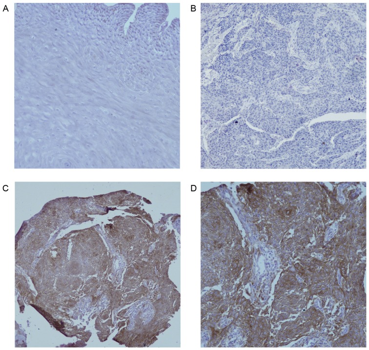
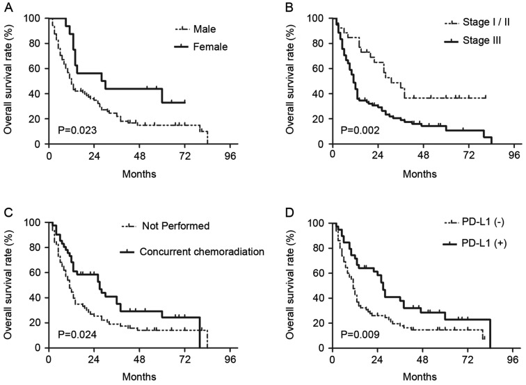
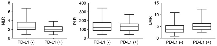
Immunotherapy with anti-programmed cell death protein 1 or programmed death ligand 1 (PD-L1) agents has demonstrated promising efficacy for the treatment of various types of malignancies. However, the role of PD-L1 as a tumor prognostic marker remains poorly understood. In the present study, the prognostic value of PD-L1 expression in esophageal carcinoma (EC) following definitive chemoradiotherapy (CRT) was investigated, and its associations with three systemic inflammation biomarkers, neutrophil-to-lymphocyte ratio (NLR), platelet-to-lymphocyte ratio (PLR) and lymphocyte-to-monocyte ratio (LMR) were further explored. A total of 104 patients with non-metastatic EC, who underwent definitive CRT between January 2009 and December 2012, were retrospectively analyzed. The expression of PD-L1 was examined by immunohistochemistry and the impact of PD-L1 expression level on overall survival (OS) was assessed. Furthermore, pretreatment neutrophil, lymphocyte, platelet and monocyte counts were obtained from routine blood tests to calculate the NLR, PLR and LMR. PD-L1 was overexpressed in EC compared with normal esophageal epithelium, with a positive expression rate of 37.5%. Additionally, patients with positive PD-L1 expression had a lower NLR than those with negative PD-L1 expression (P=0.001). On multivariate analysis, the positive staining of PD-L1 was significantly associated with improved OS (HR, 0.6; 95% CI, 0.372-0.965; P=0.035). Kaplan-Meier survival analysis showed a similar result (P=0.009). Additionally, sex (HR, 0.449; 95% CI, 0.229-0.880; P=0.020), clinical stage III (HR, 2.471; 95% CI, 1.171-5.212; P=0.018), and receipt of concurrent chemoradiation (HR, 0.590; 95% CI, 0.368-0.945; P=0.028) were all independent prognostic factors in EC treated with definitive CRT. The correlation of NLR with PD-L1 expression validated the relevance of immunity and inflammation. In summary, the present study demonstrated that positive PD-L1 expression is associated with improved survival in patients with EC treated with radical CRT, indicating that PD-L1 is a promising prognostic marker.
使用抗程序性细胞死亡蛋白1或程序性死亡配体1(PD-L1)药物进行免疫治疗已在各种类型恶性肿瘤的治疗中显示出有前景的疗效。然而,PD-L1作为肿瘤预后标志物的作用仍知之甚少。在本研究中,我们调查了PD-L1表达在食管癌(EC)根治性放化疗(CRT)后的预后价值,并进一步探讨了其与三种全身炎症生物标志物,即中性粒细胞与淋巴细胞比值(NLR)、血小板与淋巴细胞比值(PLR)和淋巴细胞与单核细胞比值(LMR)之间的关联。我们对2009年1月至2012年12月期间接受根治性CRT的104例非转移性EC患者进行了回顾性分析。通过免疫组织化学检测PD-L1的表达,并评估PD-L1表达水平对总生存期(OS)的影响。此外,从常规血液检查中获取治疗前中性粒细胞、淋巴细胞、血小板和单核细胞计数,以计算NLR、PLR和LMR。与正常食管上皮相比,PD-L1在EC中过表达,阳性表达率为37.5%。此外,PD-L1表达阳性的患者NLR低于PD-L1表达阴性的患者(P = 0.001)。多因素分析显示,PD-L1阳性染色与OS改善显著相关(HR,0.6;95%CI,0.372 - 0.965;P = 0.035)。Kaplan-Meier生存分析显示了类似结果(P = 0.009)。此外,性别(HR,0.449;95%CI,0.229 - 0.880;P = 0.020)、临床III期(HR,2.471;95%CI,1.171 - 5.212;P = 0.018)以及接受同步放化疗(HR,0.590;95%CI,0.368 - 0.945;P = 0.028)均为根治性CRT治疗的EC患者的独立预后因素。NLR与PD-L1表达的相关性验证了免疫与炎症的相关性。总之,本研究表明,根治性CRT治疗的EC患者中,PD-L1阳性表达与生存期改善相关,表明PD-L1是一个有前景的预后标志物。