(ret) Robert Koch Institute, Centre for Biological Threats and Special Pathogens, ZBS 4: Advanced Light and Electron Microscopy, Seestrasse 10, D-13353 Berlin, Germany.
(ret) University of Newcastle upon Tyne, Burnfoot, Stocksfield, Northumberland, NE43 7TN, UK.
Viruses. 2018 Mar 22;10(4):142. doi: 10.3390/v10040142.
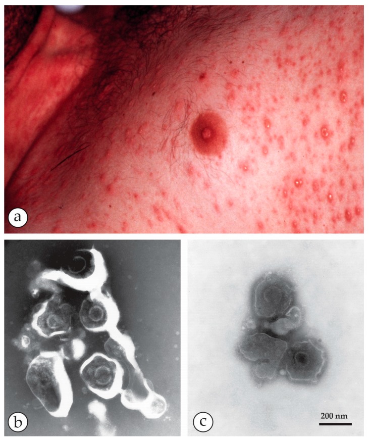
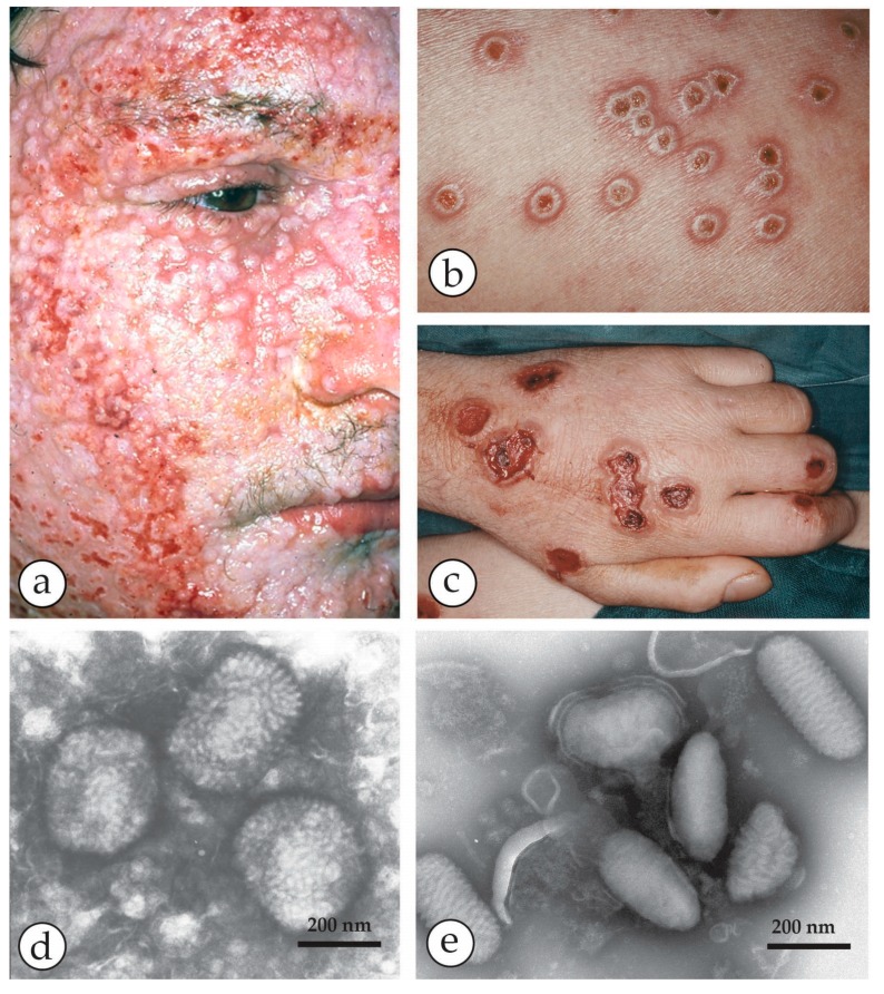
Diagnostic electron microscopy (DEM) was an essential component of viral diagnosis until the development of highly sensitive nucleic acid amplification techniques (NAT). The simple negative staining technique of DEM was applied widely to smallpox diagnosis until the world-wide eradication of the human-specific pathogen in 1980. Since then, the threat of smallpox re-emerging through laboratory escape, molecular manipulation, synthetic biology or bioterrorism has not totally disappeared and would be a major problem in an unvaccinated population. Other animal poxviruses may also emerge as human pathogens. With its rapid results (only a few minutes after arrival of the specimen), no requirement for specific reagents and its "open view", DEM remains an important component of virus diagnosis, particularly because it can easily and reliably distinguish smallpox virus or any other member of the orthopoxvirus (OPV) genus from parapoxviruses (PPV) and the far more common and less serious herpesviruses (herpes simplex and varicella zoster). Preparation, enrichment, examination, internal standards and suitable organisations are discussed to make clear its continuing value as a diagnostic technique.
诊断电子显微镜(DEM)是病毒诊断的重要组成部分,直到高度敏感的核酸扩增技术(NAT)的发展。DEM 的简单负染色技术广泛应用于天花诊断,直到 1980 年世界范围内消灭了这种特定于人类的病原体。此后,天花通过实验室逃逸、分子操作、合成生物学或生物恐怖主义重新出现的威胁并没有完全消失,而且在未接种疫苗的人群中会成为一个主要问题。其他动物痘病毒也可能成为人类病原体。由于其快速的结果(标本到达后仅几分钟)、不需要特定试剂以及其“开放式观察”,DEM 仍然是病毒诊断的重要组成部分,特别是因为它可以轻松可靠地区分天花病毒或任何其他正痘病毒(OPV)属成员与副痘病毒(PPV)和更为常见且不太严重的疱疹病毒(单纯疱疹和带状疱疹)。讨论了制备、富集、检查、内标和合适的组织,以明确其作为诊断技术的持续价值。