Hashimoto Kazuhiko, Oda Yutaka, Nakagawa Koichi, Ikeda Terumasa, Ohtani Kazuhiro, Akagi Masao
Kindai University Hospital.
Eur J Histochem. 2018 Jan 22;62(1):2847. doi: 10.4081/ejh.2018.2847.
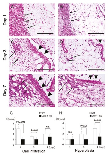
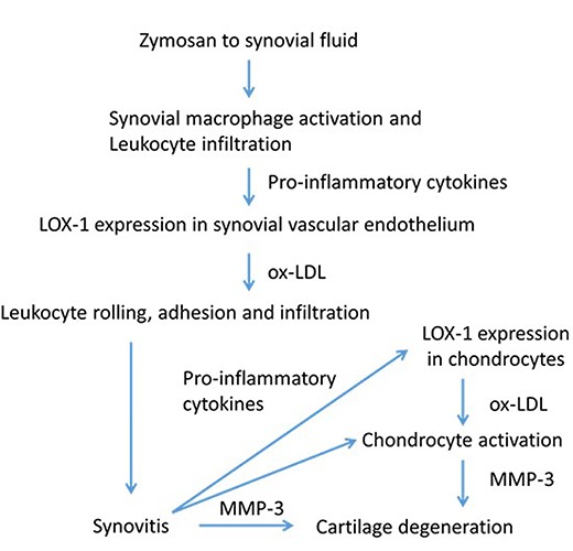
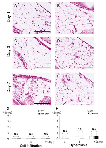
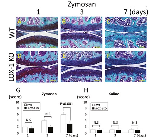
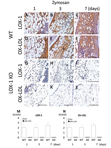
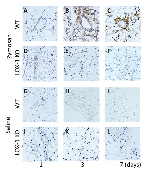
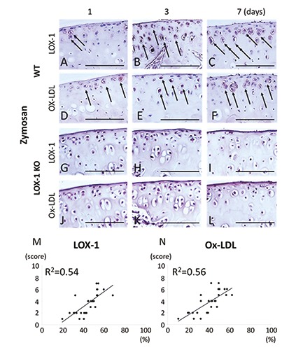
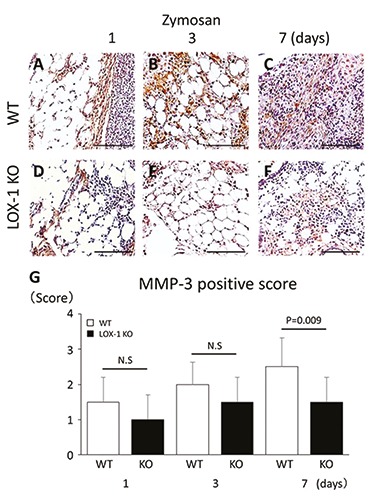
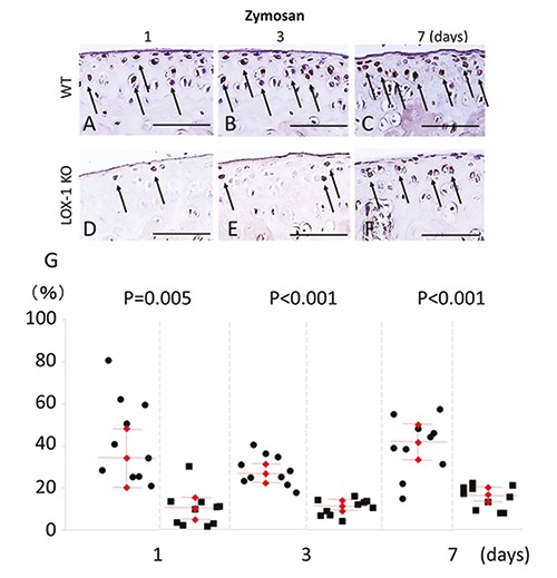
Recent data suggest that the lectin-like oxidized low-density lipoprotein (ox-LDL) receptor-1 (LOX-1)/ox-LDL system may be involved in the pathogenesis of arthritis. We aimed to demonstrate the roles of the LOX-1/ox-LDL system in arthritis development by using LOX-1 knockout (KO) mice. Arthritis was induced in the right knees of C57Bl/6 wild-type (WT) and LOX-1 KO mice via zymosan injection. Saline was injected in the left knees. Arthritis development was evaluated using inflammatory cell infiltration, synovial hyperplasia, and cartilage degeneration scores at 1, 3, and 7 days after administration. LOX-1, ox-LDL, and matrix metalloproteinase-3 (MMP-3) expression in the synovial cells and chondrocytes was evaluated by immunohistochemistry. The LOX-1, ox-LDL, and MMP-3 expression levels in synovial cells were scored on a grading scale. The positive cell rate of LOX-1, ox-LDL, and MMP-3 in chondrocytes was measured. The correlation between the positive cell rate of LOX-1 or ox-LDL and the cartilage degeneration score was also examined. Inflammatory cell infiltration, synovial hyperplasia, and cartilage degeneration were significantly reduced in the LOX-1 KOmice with zymosan-induced arthritis (ZIA) compared to WT mice with ZIA. In the saline-injected knees, no apparent arthritic changes were observed. LOX-1 and ox-LDL expression in synovial cells and chondrocytes were detected in the knees of WT mice with ZIA. No LOX-1 and ox-LDL expression was detected in the knees of LOX-1 KOmice with ZIA or the saline-injected knees of both mice. MMP-3 expression in the synovial cells and chondrocytes was also detected in knees of both mice with ZIA, and was significantly less in the LOX-1 KO mice than in WT mice. The positive cell rate of LOX-1 or ox-LDL and the cartilage degeneration score showed a positive correlation. Our data show the involvement of the LOX-1/ox-LDL system in murine ZIA development. LOX-1-positive synovial cells and chondrocytes are potential therapeutic targets for arthritis prevention.
近期数据表明,凝集素样氧化型低密度脂蛋白(ox-LDL)受体-1(LOX-1)/ox-LDL系统可能参与了关节炎的发病机制。我们旨在通过使用LOX-1基因敲除(KO)小鼠来证明LOX-1/ox-LDL系统在关节炎发展中的作用。通过向C57Bl/6野生型(WT)和LOX-1 KO小鼠的右膝注射酵母聚糖诱导关节炎。左膝注射生理盐水。在给药后1、3和7天,使用炎症细胞浸润、滑膜增生和软骨退变评分来评估关节炎的发展。通过免疫组织化学评估滑膜细胞和软骨细胞中LOX-1、ox-LDL和基质金属蛋白酶-3(MMP-3)的表达。对滑膜细胞中LOX-1、ox-LDL和MMP-3的表达水平进行分级评分。测量软骨细胞中LOX-1、ox-LDL和MMP-3的阳性细胞率。还检查了LOX-1或ox-LDL的阳性细胞率与软骨退变评分之间的相关性。与患有酵母聚糖诱导关节炎(ZIA)的WT小鼠相比,患有ZIA的LOX-1 KO小鼠的炎症细胞浸润、滑膜增生和软骨退变明显减少。在注射生理盐水的膝盖中,未观察到明显的关节炎变化。在患有ZIA的WT小鼠的膝盖中检测到滑膜细胞和软骨细胞中LOX-1和ox-LDL的表达。在患有ZIA的LOX-1 KO小鼠的膝盖或两种小鼠注射生理盐水的膝盖中未检测到LOX-1和ox-LDL的表达。在患有ZIA的两种小鼠的膝盖中也检测到滑膜细胞和软骨细胞中MMP-3的表达,并且在LOX-1 KO小鼠中明显低于WT小鼠。LOX-1或ox-LDL的阳性细胞率与软骨退变评分呈正相关。我们的数据表明LOX-1/ox-LDL系统参与了小鼠ZIA的发展。LOX-1阳性的滑膜细胞和软骨细胞是预防关节炎的潜在治疗靶点。