Department of Radiology, Affiliated Hospital of Nanjing University of Chinese Medicine, Nanjing 210029, China; Division of Nephrology, Zhongshan Hospital Fudan University, Shanghai 200032, China.
Department of Radiology, Affiliated Hospital of Nanjing University of Chinese Medicine, Nanjing 210029, China.
EBioMedicine. 2018 Apr;30:129-137. doi: 10.1016/j.ebiom.2018.03.008. Epub 2018 Mar 15.
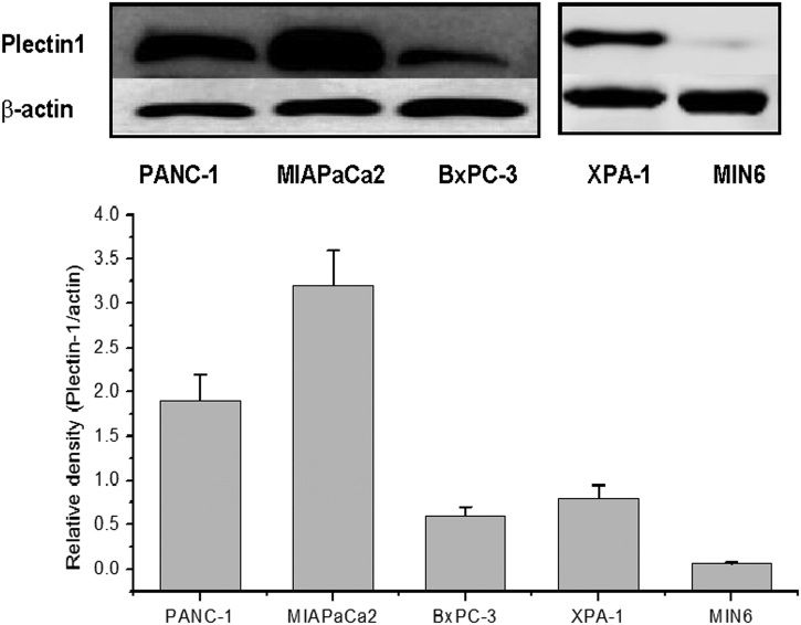
Biomarker-targeted molecular imaging holds promise for early detection of pancreatic cancer. The aim of this study was to design and evaluate a plectin-1 targeted multi-functional nanoparticle probe for pancreatic cancer imaging.
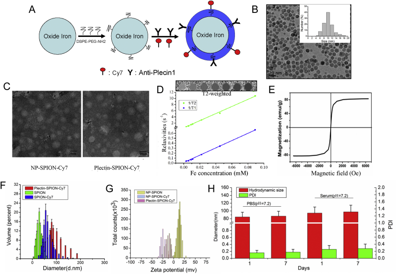
1,2-Distearoyl-sn-glycero-3-phosphoethanolamine-N-amino(polyethylene glycol) (DSPE-PEG-NH2)-modified superparamagnetic iron oxide (FeO) nanoparticles (SPION) were conjugated with plectin-1 antibody and/or Cy7 to create the multi-functional targeted nanoparticle targeted probe (Plectin-SPION-Cy7) or non-targeted probe (SPION-Cy7). Pancreatic carcinoma cell lines expressing plectin-1 were cultured with the targeted or control probes and then were imaged using confocal laser scanning microscopy and magnetic resonance imaging (MRI). Accumulations of the nanoparticles in pancreatic tumor xenografted mice were determined by MRI and fluorescence imaging.
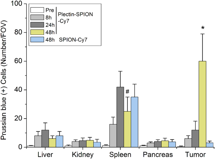
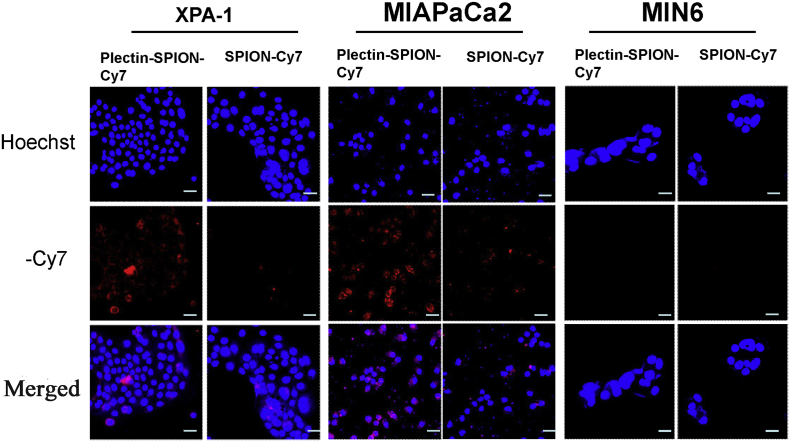
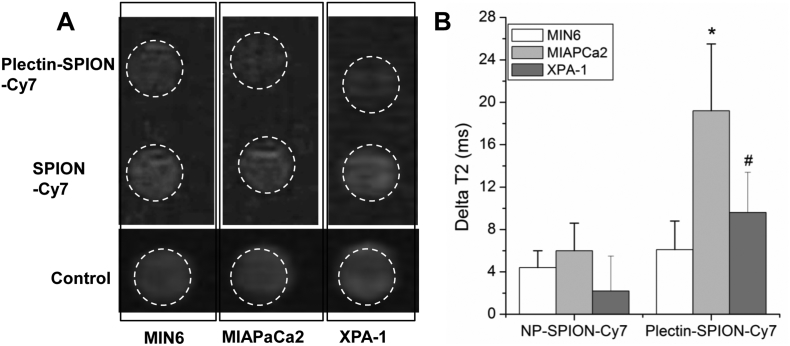
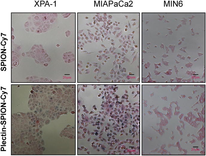
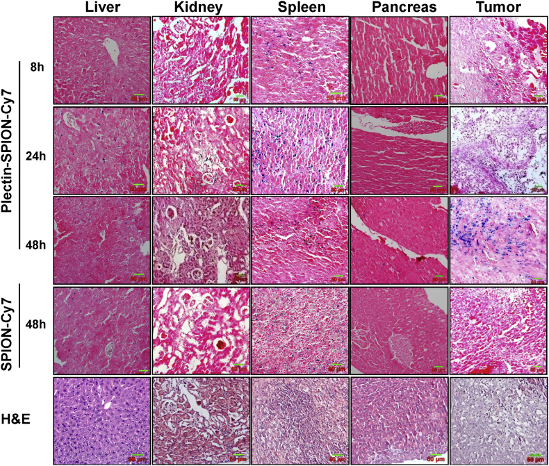
In vitro optical imaging and MRI showed that the targeted nanoparticles were highly accumulated in MIAPaCa2 and XPA-1 carcinoma cells but not in non-carcinoma MIN6 cells, which was further confirmed by Prussian blue staining. In vivo MRI showed a significant T2 signal reduction. Prussian blue staining further confirmed that the plectin-1 targeted nanoparticles were highly accumulated in the tumor mass but not in normal pancreatic tissues, or in the liver and kidney, and few nanoparticles were observed in the tumors of mice injected with SPION-Cy7.
Our data demonstrate that plectin-1 targeted fluorescence and MR dual-functional nanoparticle can visualize pancreatic cancer, and it has great potential to be used with various imaging devices for pancreatic cancer detection.
生物标志物靶向分子成像有望实现胰腺癌的早期检测。本研究旨在设计并评估一种靶向黏着斑蛋白-1 的多功能纳米颗粒探针,用于胰腺癌成像。
将 1,2-二硬脂酰-sn-甘油-3-磷酸乙醇胺-N-氨基(聚乙二醇)(DSPE-PEG-NH2)修饰的超顺磁性氧化铁(FeO)纳米颗粒(SPION)与黏着斑蛋白-1 抗体和/或 Cy7 偶联,以创建多功能靶向纳米颗粒靶向探针(Plectin-SPION-Cy7)或非靶向探针(SPION-Cy7)。表达黏着斑蛋白-1 的胰腺癌细胞系与靶向或对照探针孵育,然后使用共聚焦激光扫描显微镜和磁共振成像(MRI)进行成像。通过 MRI 和荧光成像确定胰腺肿瘤异种移植小鼠中纳米颗粒的积累。
体外光学成像和 MRI 显示,靶向纳米颗粒在 MIAPaCa2 和 XPA-1 癌细胞中高度积累,但在非癌细胞 MIN6 中则没有,普鲁士蓝染色进一步证实了这一点。体内 MRI 显示 T2 信号显著降低。普鲁士蓝染色进一步证实,黏着斑蛋白-1 靶向纳米颗粒在肿瘤组织中高度积累,但在正常胰腺组织、肝脏和肾脏中则没有,在注射 SPION-Cy7 的小鼠肿瘤中观察到的纳米颗粒很少。
我们的数据表明,黏着斑蛋白-1 靶向荧光和磁共振双功能纳米颗粒可以可视化胰腺癌,并且具有与各种成像设备一起用于胰腺癌检测的巨大潜力。