Mohammadi Tooba, Gheybalizadeh Hadi, Rahimpour Elaheh, Soleymani Jafar, Shafiei-Irannejad Vahid
Student Research Committee, Urmia University of Medical Sciences, Urmia, Iran.
Liver and Gastrointestinal Diseases Research Center, Tabriz University of Medical Sciences, Tabriz, Iran.
Heliyon. 2024 Dec 31;11(1):e41566. doi: 10.1016/j.heliyon.2024.e41566. eCollection 2025 Jan 15.
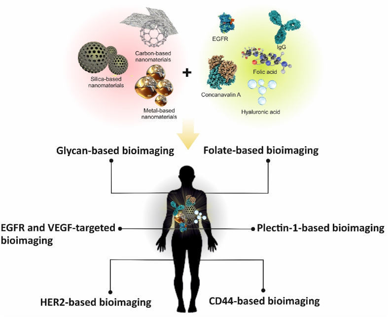
The investigation of changes in the membrane of cancer cells holds great potential for biomedical applications. Malignant cells exhibit overexpression of receptors, which can be used for targeted drug delivery, therapy, and bioimaging. Targeted bioimaging is one the most accurate imaging methods with a non-invasive nature, allowing for localization of the malignant cell without disrupting cellular integrity. Also, bioimaging has the potential to enhance the quality of established imaging techniques like magnetic resonance imaging (MRI). The utilization of nanoparticles in targeted bioimaging enhances the imaging quality and efficiency. Biocompatible nanoparticles can easily penetrate cell membranes, while they can be readily functionalized on their surfaces toward cell receptors. This study reviews reports on the application of new advanced photoluminescent materials for targeted bioimaging using the cell membrane receptors. Also, the limitations and advantages of the application of nanoparticles have been reviewed along with the clinical consideration of their uses in bioimaging.
对癌细胞膜变化的研究在生物医学应用方面具有巨大潜力。恶性细胞表现出受体的过度表达,这可用于靶向药物递送、治疗和生物成像。靶向生物成像是最精确的成像方法之一,具有非侵入性,能够在不破坏细胞完整性的情况下定位恶性细胞。此外,生物成像有潜力提高如磁共振成像(MRI)等现有成像技术的质量。纳米颗粒在靶向生物成像中的应用提高了成像质量和效率。生物相容性纳米颗粒能够轻松穿透细胞膜,同时它们可以很容易地在其表面进行功能化以靶向细胞受体。本研究综述了关于使用细胞膜受体进行靶向生物成像的新型先进光致发光材料应用的报告。此外,还综述了纳米颗粒应用的局限性和优势以及它们在生物成像中应用的临床考量。