School of Biomedical Engineering and Imaging Sciences, 4th Floor, North Wing, St Thomas' Hospital, King's College London, London, UK.
Department of Cardiology, Guy's and St Thomas' Hospital NHS Foundation Trust, London, UK.
Europace. 2018 Nov 1;20(11):1721-1732. doi: 10.1093/europace/euy040.
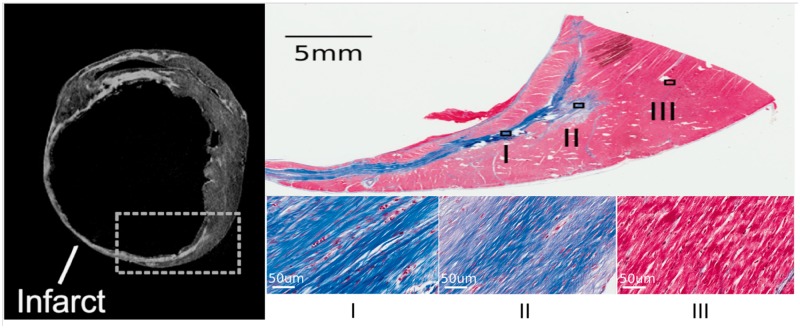
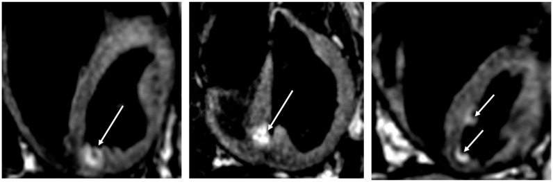
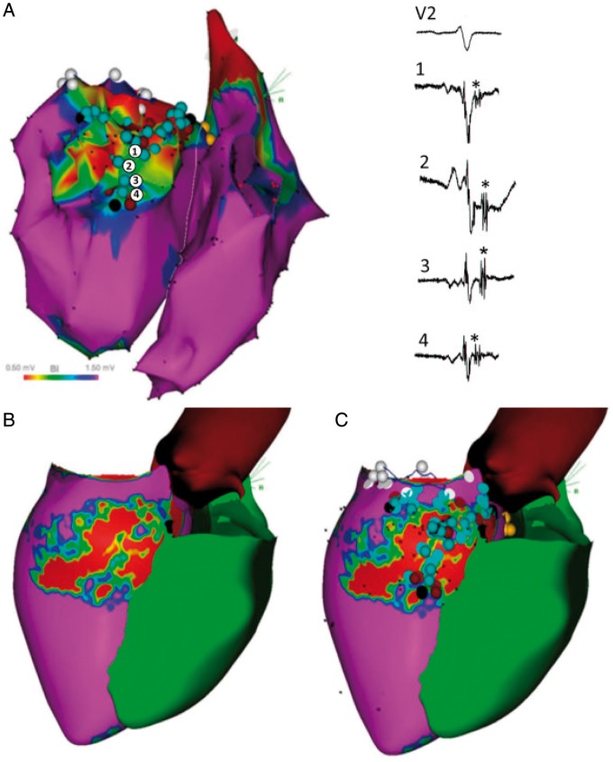
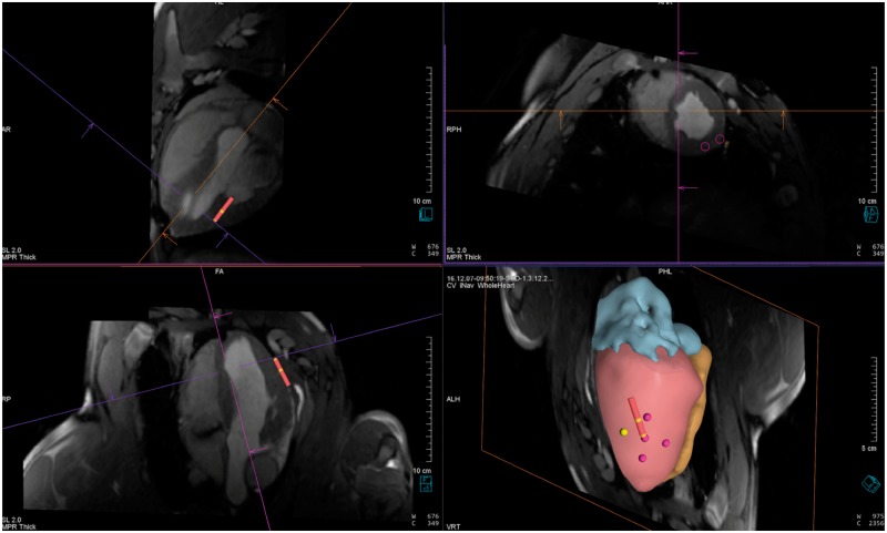
Catheter ablation has an important role in the management of patients with ventricular tachycardia (VT) but is limited by modest long-term success rates. Magnetic resonance imaging (MRI) can provide valuable anatomic and functional information as well as potentially improve identification of target sites for ablation. A major limitation of current MRI protocols is the spatial resolution required to identify the areas of tissue responsible for VT but recent developments have led to new strategies which may improve substrate assessment. Potential ways in which detailed information gained from MRI may be utilized during electrophysiology procedures include image integration or performing a procedure under real-time MRI guidance. Image integration allows pre-procedural magnetic resonance (MR) images to be registered with electroanatomical maps to help guide VT ablation and has shown promise in preliminary studies. However, multiple errors can arise during this process due to the registration technique used, changes in ventricular geometry between the time of MRI and the ablation procedure, respiratory and cardiac motion. As isthmus sites may only be a few millimetres wide, reducing these errors may be critical to improve outcomes in VT ablation. Real-time MR-guided intervention has emerged as an alternative solution to address the limitations of pre-acquired imaging to guide ablation. There is now a growing body of literature describing the feasibility, techniques, and potential applications of real-time MR-guided electrophysiology. We review whether real-time MR-guided intervention could be applied in the setting of VT ablation and the potential challenges that need to be overcome.
导管消融在室性心动过速(VT)患者的治疗中具有重要作用,但长期成功率有限。磁共振成像(MRI)可以提供有价值的解剖和功能信息,并可能有助于确定消融的目标部位。目前 MRI 方案的一个主要限制是识别导致 VT 的组织区域所需的空间分辨率,但最近的发展导致了新的策略,这些策略可能改善底物评估。从 MRI 获得的详细信息在电生理程序中可能被利用的潜在方法包括图像整合或在实时 MRI 引导下进行程序。图像整合允许将术前磁共振(MR)图像与电解剖图进行配准,以帮助指导 VT 消融,并在初步研究中显示出前景。然而,由于所使用的配准技术、MRI 时间和消融程序之间心室几何形状的变化、呼吸和心脏运动,在这个过程中可能会出现多个错误。由于峡部部位可能只有几毫米宽,因此减少这些错误对于改善 VT 消融的结果可能至关重要。实时 MR 引导干预已成为解决预采集图像引导消融的局限性的替代解决方案。现在有越来越多的文献描述了实时 MR 引导电生理的可行性、技术和潜在应用。我们回顾了实时 MR 引导干预是否可以应用于 VT 消融中,以及需要克服的潜在挑战。