Burger Janneke C, Hopman Luuk H G A, Kemme Michiel J B, Hoeksema Wiert, Takx Richard A P, Figueras I Ventura Rosa M, Campos Fernando O, Plank Gernot, Planken R Nils, Allaart Cornelis P, van Halm Vokko P, Postema Pieter G, Götte Marco J W, Bishop Martin J, Bhagirath Pranav
Department of Cardiology, Amsterdam University Medical Center, Amsterdam, The Netherlands.
Department of Radiology and Nuclear Medicine, Amsterdam University Medical Center, Amsterdam, The Netherlands.
Heart Rhythm O2. 2024 Jul 5;5(8):561-572. doi: 10.1016/j.hroo.2024.07.001. eCollection 2024 Aug.
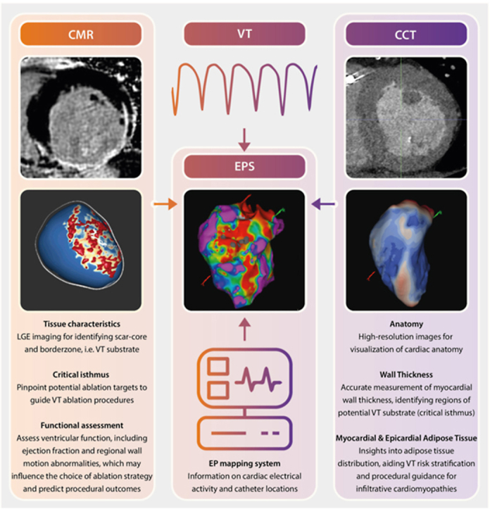
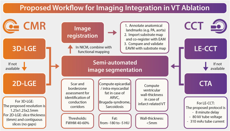
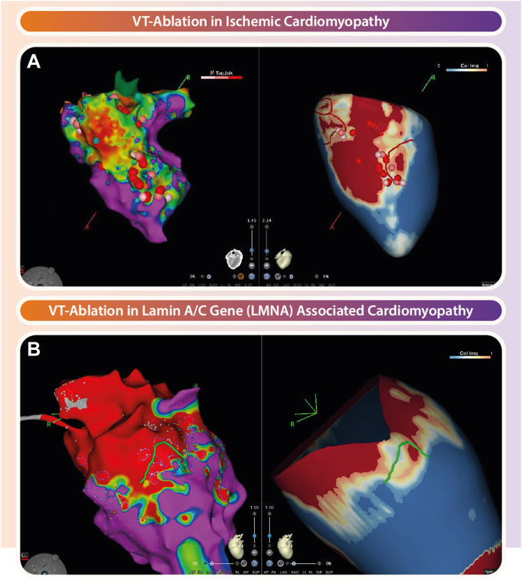
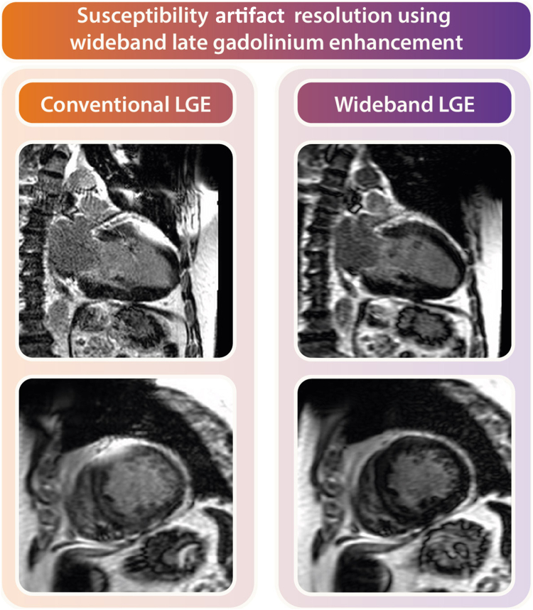
Ventricular tachycardia (VT) is a life-threatening heart rhythm and has long posed a complex challenge in the field of cardiology. Recent developments in advanced imaging modalities have aimed to improve comprehension of underlying arrhythmic substrate for VT. To this extent, high-resolution cardiac magnetic resonance (CMR) and cardiac computed tomography (CCT) have emerged as tools for accurately visualizing and characterizing scar tissue, fibrosis, and other critical structural abnormalities within the heart, providing novel insights into VT triggers and substrate. However, clinical implementation of knowledge derived from these advanced imaging techniques in improving VT treatment and guiding invasive therapeutic strategies continues to pose significant challenges. A pivotal concern lies in the absence of standardized imaging protocols and analysis methodologies, resulting in a large variance in data quality and consistency. Furthermore, the clinical significance and outcomes associated with VT substrate characterization through CMR and CCT remain dynamic and subject to ongoing evolution. This highlights the need for refinement of these techniques before their reliable integration into routine patient care can be realized. The primary objectives of this study are twofold: firstly, to provide a comprehensive overview of the studies conducted over the last 15 years, summarizing the current available literature on imaging-based assessment of VT substrate. Secondly, to critically analyze and evaluate the selected studies, with the aim of providing valuable insights that can inform current clinical practice and future research.
室性心动过速(VT)是一种危及生命的心律,长期以来一直是心脏病学领域的一个复杂挑战。先进成像技术的最新发展旨在提高对VT潜在心律失常基质的理解。在这方面,高分辨率心脏磁共振成像(CMR)和心脏计算机断层扫描(CCT)已成为准确可视化和表征心脏内瘢痕组织、纤维化及其他关键结构异常的工具,为VT的触发因素和基质提供了新的见解。然而,将这些先进成像技术所获得的知识应用于改善VT治疗和指导侵入性治疗策略的临床实践仍面临重大挑战。一个关键问题在于缺乏标准化的成像方案和分析方法,导致数据质量和一致性存在很大差异。此外,通过CMR和CCT对VT基质进行表征的临床意义和结果仍在动态变化且不断演变。这凸显了在将这些技术可靠地整合到常规患者护理之前对其进行完善的必要性。本研究的主要目标有两个:第一,全面概述过去15年进行的研究,总结目前关于基于成像的VT基质评估的现有文献。第二,批判性地分析和评估所选研究,旨在提供有价值的见解,为当前临床实践和未来研究提供参考。