Center for Cellular and Molecular Engineering, Department of Orthopaedic Surgery, University of Pittsburgh School of Medicine, 450 Technology Drive, Room 221, Pittsburgh, PA, 15219, USA.
Department of Bioengineering, Swanson School of Engineering, University of Pittsburgh, Pittsburgh, PA, 15219, USA.
Stem Cell Res Ther. 2018 Apr 3;9(1):86. doi: 10.1186/s13287-018-0830-4.
Adult mesenchymal stem cells (MSCs) are an important resource for tissue growth, repair, and regeneration. To utilize MSCs more effectively, a clear understanding of how they react to environmental cues is essential. Currently, relatively little is known about how the composition of the plasma membranes affects stem cell phenotype and properties. The presence of lipid molecules, including cholesterol in particular, in the plasma membrane plays a crucial role in regulating a variety of physiological processes in cells. In this study, we examined the effects of perturbations in cholesterol/caveolin-1 (CAV-1)/caveolae homeostasis on the membrane properties and adhesive characteristics of MSCs. Findings from this study will contribute to the understanding of how cholesterol/CAV-1/caveolae regulates aspects of the cell membrane important to cell adhesion, substrate sensing, and microenvironment interaction.
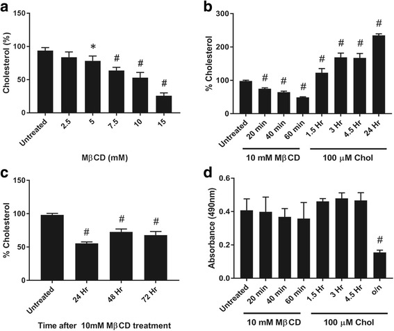
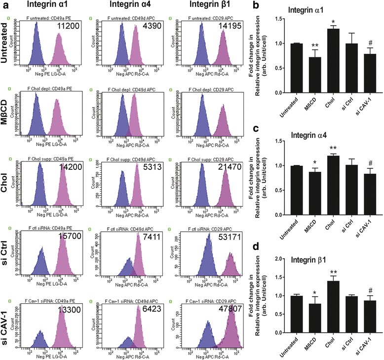
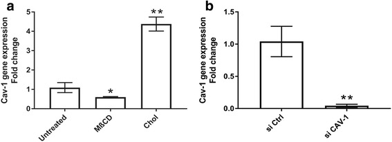
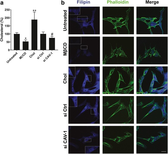
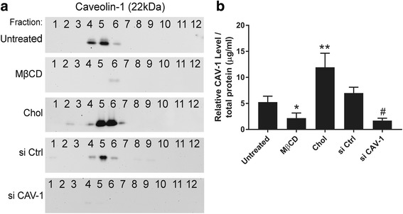
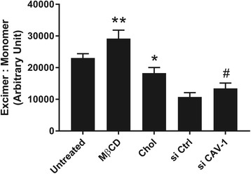
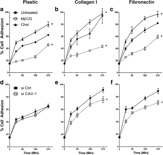
We generated five experimental MSC groups: 1) untreated MSCs; 2) cholesterol-depleted MSCs; 3) cholesterol-supplemented MSCs; 4) MSCs transfected with control, nonspecific small interfering (si)RNA; and 5) MSCs transfected with CAV-1 siRNA. Each cell group was analyzed for perturbation of cholesterol status and CAV-1 expression by performing Amplex Red cholesterol assay, filipin fluorescence staining, and real-time polymerase chain reaction (PCR). The membrane fluidity in the five experimental cell groups were measured using pyrene fluorescence probe staining followed by FACS analysis. Cell adhesion to collagen and fibronectin as well as cell surface integrin expression were examined.
Cholesterol supplementation to MSCs increased membrane cholesterol, and resulted in decreased membrane fluidity and localization of elevated numbers of caveolae and CAV-1 to the cell membrane. These cells showed increased expression of α1, α4, and β1 integrins, and exhibited higher adhesion rates to fibronectin and collagen. Conversely, knockdown of CAV-1 expression or cholesterol depletion on MSCs caused a parallel decrease in caveolae content and an increase in membrane fluidity due to decreased delivery of cholesterol to the cell membrane. Cells with depleted CAV-1 expression showed decreased cell surface integrin expression and slower adhesion to different substrates.
Our results demonstrate that perturbations in cholesterol/CAV-1 levels significantly affect the membrane properties of MSCs. These findings suggest that modification of membrane cholesterol and/or CAV-1 and caveolae may be used to manipulate the biological activities of MSCs.
成体间充质干细胞(MSCs)是组织生长、修复和再生的重要资源。为了更有效地利用 MSCs,必须清楚地了解它们对环境线索的反应。目前,关于质膜的组成如何影响干细胞表型和特性的了解相对较少。质膜中脂质分子(包括胆固醇)的存在在调节细胞内的各种生理过程中起着至关重要的作用。在这项研究中,我们研究了胆固醇/窖蛋白-1(CAV-1)/小窝内陷稳态的干扰对 MSCs 膜特性和黏附特性的影响。本研究的结果将有助于了解胆固醇/CAV-1/小窝内陷如何调节与细胞黏附、底物感知和微环境相互作用有关的细胞膜的各个方面。
我们生成了五个实验 MSC 组:1)未处理的 MSC;2)胆固醇耗尽的 MSC;3)胆固醇补充的 MSC;4)转染了对照、非特异性小干扰(si)RNA 的 MSC;5)转染了 CAV-1 siRNA 的 MSC。通过 Amplex Red 胆固醇测定、佛波醇荧光染色和实时聚合酶链反应(PCR)分析,对每个细胞组进行胆固醇状态和 CAV-1 表达的扰动分析。使用芘荧光探针染色后通过流式细胞术分析,测量五个实验细胞组的膜流动性。检测细胞对胶原蛋白和纤维连接蛋白的黏附以及细胞表面整联蛋白的表达。
向 MSC 中补充胆固醇会增加质膜中的胆固醇,导致质膜流动性降低,并且大量 caveolae 和 CAV-1 定位于质膜。这些细胞表现出更高水平的α1、α4 和β1 整联蛋白表达,并表现出更高的纤维连接蛋白和胶原蛋白黏附率。相反,敲低 MSC 中的 CAV-1 表达或耗尽胆固醇会导致 caveolae 含量平行减少,并且由于胆固醇向质膜的输送减少而导致膜流动性增加。耗尽 CAV-1 表达的细胞表现出更低的细胞表面整联蛋白表达和对不同底物的更慢黏附。
我们的结果表明,胆固醇/CAV-1 水平的扰动会显著影响 MSCs 的膜特性。这些发现表明,膜胆固醇和/或 CAV-1 和 caveolae 的修饰可用于操纵 MSCs 的生物学活性。