Department of Medicine, Section of Hematology/Oncology, Comprehensive Cancer Center, University of Chicago Medicine, 5841 S. South Maryland Avenue, MC 2115, Chicago, IL, 60637, USA.
Department of Pathology, University of Chicago, 5841 S. Maryland Avenue, MC 2115, Chicago, IL, 60637, USA.
J Immunother Cancer. 2018 Apr 4;6(1):24. doi: 10.1186/s40425-018-0334-x.
Immunotherapies targeting the PD-1 checkpoint pathway have recently gained regulatory approval in numerous cancer types. With the widespread use of immune checkpoint therapies, varying patterns of responses and immune-related adverse events are being observed.
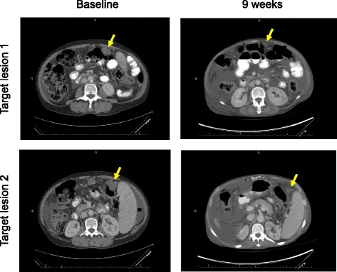
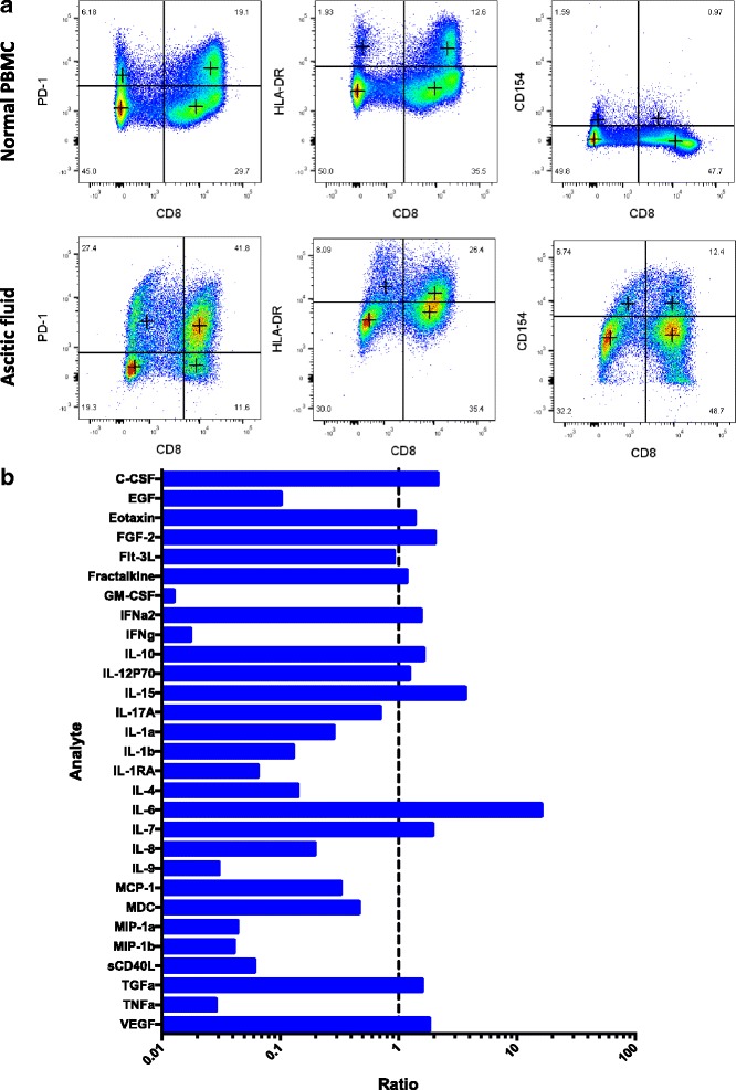
In this case, we highlight a patient who developed recurrent, large-volume ascites, while simultaneously having a 49% reduction in peritoneal tumor lesion size by RECIST criteria. Sampling of the fluid revealed high levels of IL-6 and IL-15. Cytology revealed no malignant cells on 4 separate paracenteses over a period of 6 weeks. Cell counts revealed that 45% of cells were lymphocytes, and further analysis was performed by fluorescence-activated cell sorting (FACS). The majority of lymphocytes were CD8, of which 78% were PD-1 and 43% were HLA-DR indicating an activated phenotype.
In summary, treatment with anti-PD-1 therapy may result in pseudoprogression manifested by ascitic fluid accumulation due to the influx of activated T cells. Since worsening of ascites is typically associated with disease progression, it is important to consider the possibility of pesudoprogression in such patients undergoing therapy with immune checkpoint inhibitors.
针对 PD-1 检查点通路的免疫疗法最近在多种癌症类型中获得了监管批准。随着免疫检查点疗法的广泛应用,各种不同的反应模式和免疫相关的不良反应正在被观察到。
在这个病例中,我们强调了一位患者的情况,他在 RECIST 标准下同时出现了复发性大量腹水,以及腹膜肿瘤病变大小减少了 49%。对腹水的取样显示 IL-6 和 IL-15 水平很高。细胞学检查在 6 周的 4 次单独穿刺中均未发现恶性细胞。细胞计数显示 45%的细胞为淋巴细胞,进一步通过荧光激活细胞分选(FACS)进行分析。大多数淋巴细胞为 CD8,其中 78%为 PD-1,43%为 HLA-DR,表明为激活表型。
总之,抗 PD-1 治疗可能导致假性进展,表现为由于激活的 T 细胞涌入而导致腹水积聚。由于腹水恶化通常与疾病进展相关,因此对于接受免疫检查点抑制剂治疗的此类患者,重要的是要考虑假性进展的可能性。