Gong Yi, Yin Jiayang, Tong Boding, Li Jingkun, Zeng Jiexi, Zuo Zhongkun, Ye Fei, Luo Yongheng, Xiao Jing, Xiong Wei
Department of Minimal Invasive Surgery, The Second Xiangya Hospital, Central South University, Changsha, China.
Department of Ophthalmology and Eye Research Center, The Second Xiangya Hospital, Central South University, Changsha, China.
Ther Clin Risk Manag. 2018 Mar 26;14:607-616. doi: 10.2147/TCRM.S153733. eCollection 2018.
Orbital decompression is an important surgical procedure for treatment of Graves' ophthalmopathy (GO), especially in women. It is reasonable for balanced orbital decompression of the lateral and medial wall. Various surgical approaches, including endoscopic transnasal surgery for medial wall and eye-side skin incision surgery for lateral wall, are being used nowadays, but many of them lack the validity, safety, or cosmetic effect.
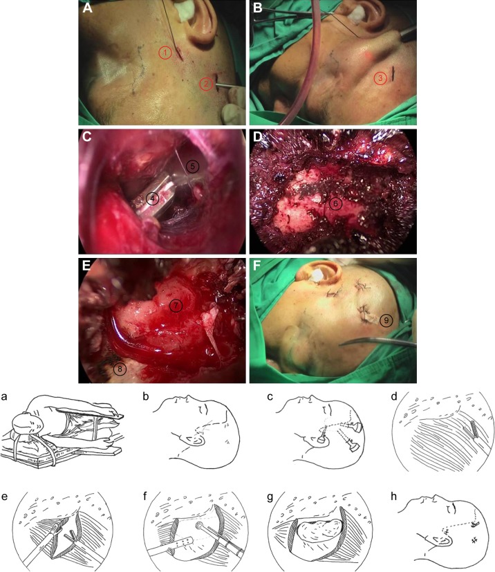
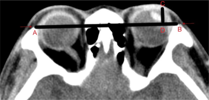
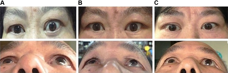
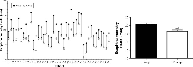
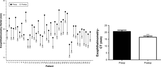
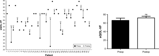
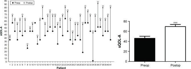
Endoscopic orbital decompression of lateral wall through hairline approach and decompression of medial wall via endoscopic transnasal surgery was done to achieve a balanced orbital decompression, aiming to improve the appearance of proptosis and create conditions for possible strabismus and eyelid surgery afterward. From January 29, 2016 to February 14, 2017, this surgery was performed on 41 orbits in 38 patients with GO, all of which were at inactive stage of disease. Just before surgery and at least 3 months after surgery, Hertel's ophthalmostatometer and computed tomography (CT) were used to check proptosis and questionnaires of GO quality of life (QOL) were completed.
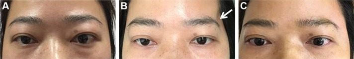
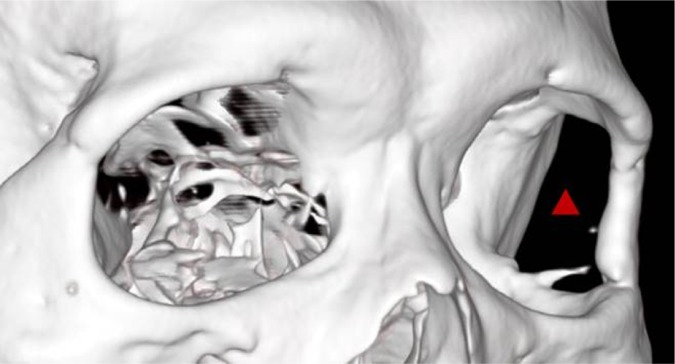
The postoperative retroversion of eyeball was 4.18±1.11 mm (Hertel's ophthalmostatometer) and 4.17±1.14 mm (CT method). The patients' QOL was significantly improved, especially the change in appearance without facial scar. The only postoperative complication was local soft tissue depression at temporal region. Obvious depression occurred in four cases (9.76%), which can be repaired by autologous fat filling.
This surgery is effective, safe, and cosmetic. Effective balanced orbital decompression can be achieved by using this original and innovative surgery method. The whole manipulation is safe and controllable under endoscope. The postoperative scar of endoscopic surgery through hairline approach is covered by hair and the anatomic structure of anterior orbit is not impacted.
眼眶减压术是治疗Graves眼病(GO)的一项重要手术,对女性患者尤为重要。进行外侧壁和内侧壁的平衡眼眶减压是合理的。目前正在使用各种手术方法,包括用于内侧壁的鼻内镜经鼻手术和用于外侧壁的眼侧皮肤切口手术,但其中许多方法缺乏有效性、安全性或美容效果。
通过发际线入路行鼻内镜下外侧壁眼眶减压术,并通过鼻内镜经鼻手术行内侧壁减压术,以实现平衡的眼眶减压,旨在改善眼球突出外观,并为后续可能的斜视和眼睑手术创造条件。2016年1月29日至2017年2月14日,对38例GO患者的41个眼眶进行了该手术,所有患者均处于疾病非活动期。术前及术后至少3个月,使用Hertel眼球突出计和计算机断层扫描(CT)检查眼球突出情况,并完成GO生活质量(QOL)问卷调查。
术后眼球后缩(Hertel眼球突出计测量)为4.18±1.11mm,(CT测量)为4.17±1.14mm。患者的生活质量得到显著改善,尤其是外观改变且无面部瘢痕。术后唯一的并发症是颞部局部软组织凹陷。4例(9.76%)出现明显凹陷,可通过自体脂肪填充修复。
该手术有效、安全且美观。采用这种原创且创新的手术方法可实现有效的平衡眼眶减压。在内窥镜下整个操作安全可控。经发际线入路的鼻内镜手术术后瘢痕被头发覆盖,不影响眶前部的解剖结构。