Fung Candice, Koussoulas Katerina, Unterweger Petra, Allen Andrew M, Bornstein Joel C, Foong Jaime P P
Department of Physiology, The University of Melbourne, Parkville, VIC, Australia.
Florey Institute of Neuroscience and Mental Health, The University of Melbourne, Parkville, VIC, Australia.
Front Physiol. 2018 Mar 21;9:260. doi: 10.3389/fphys.2018.00260. eCollection 2018.
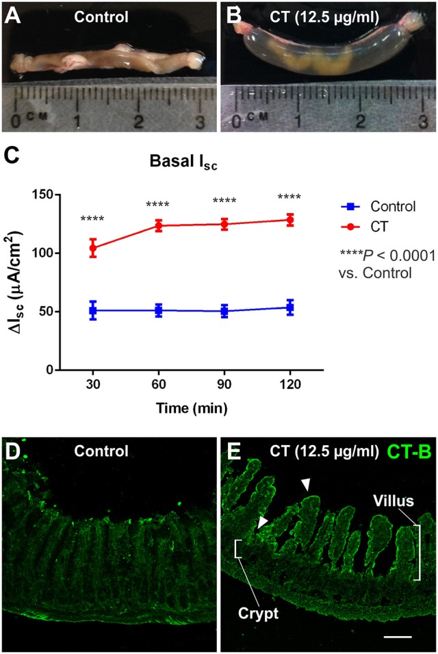
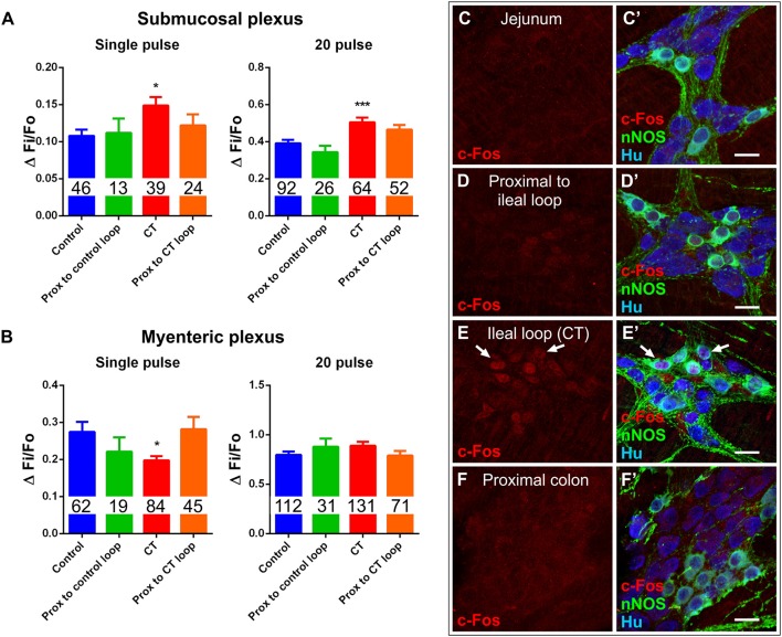
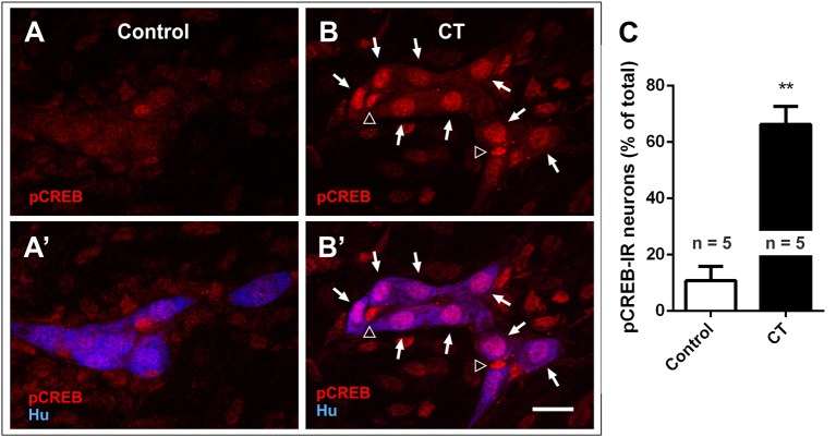
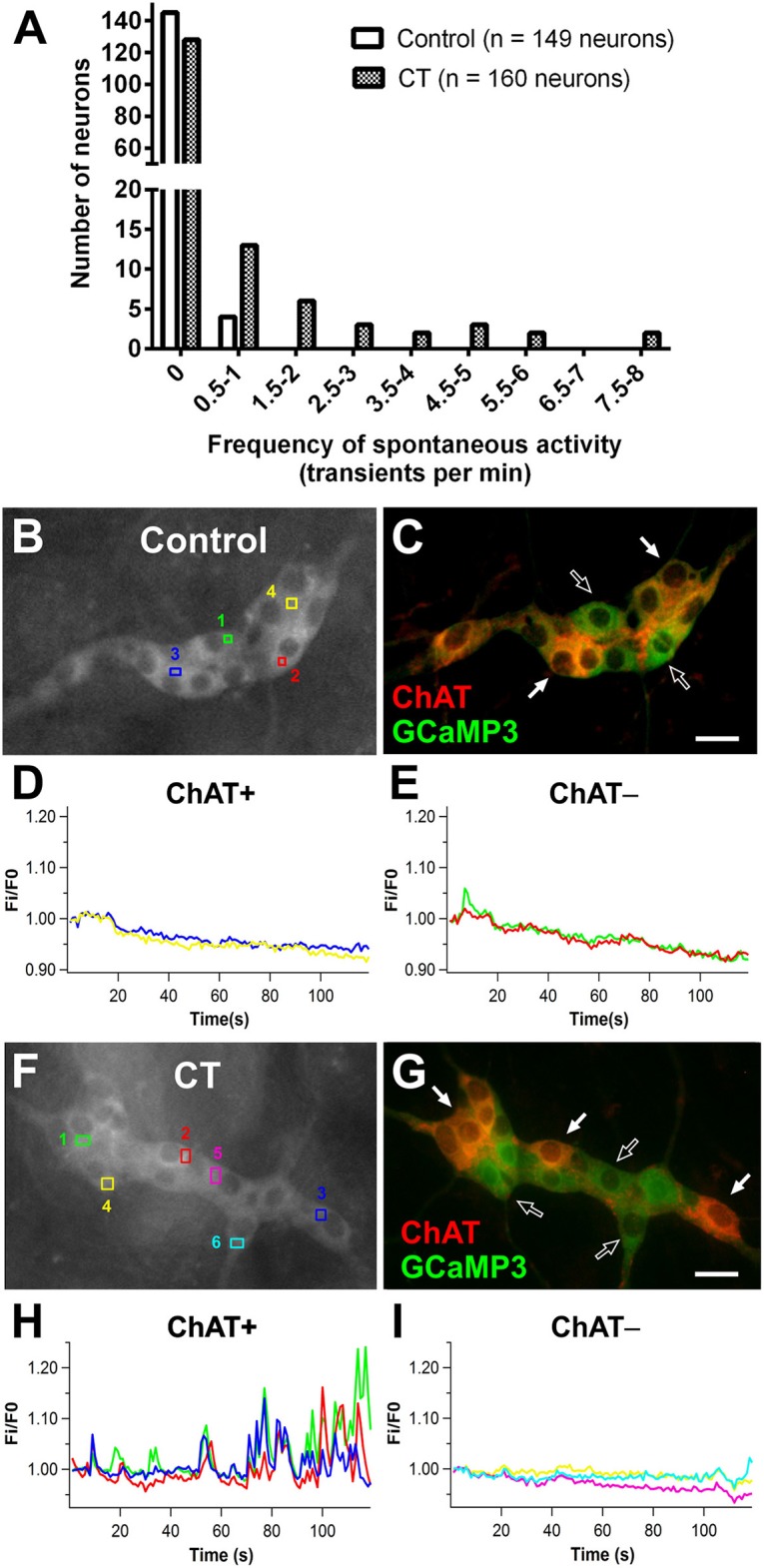
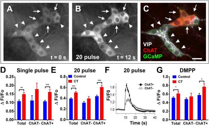
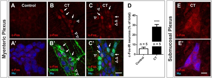
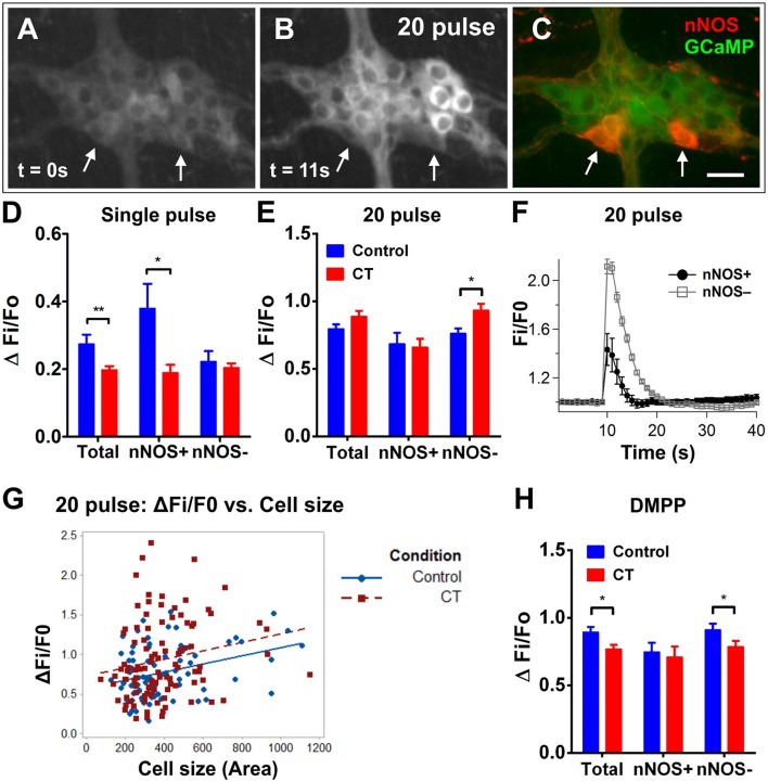
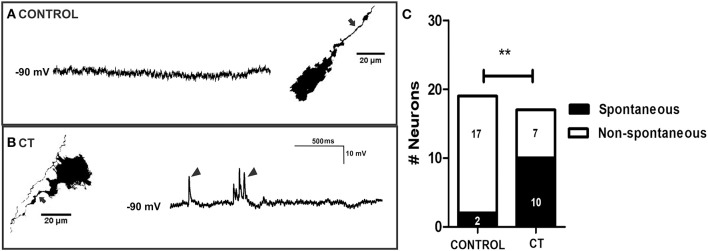
Cholera-induced hypersecretion causes dehydration and death if untreated. Cholera toxin (CT) partly acts via the enteric nervous system (ENS) and induces long-lasting changes to enteric neuronal excitability following initial exposure, but the specific circuitry involved remains unclear. We examined this by first incubating CT or saline (control) in mouse ileal loops for 3.5 h and then assessed neuronal excitability using Ca imaging and immunolabeling for the activity-dependent markers cFos and pCREB. Mice from a C57BL6 background, including -Cre;R26R- mice which express the fluorescent Ca indicator GCaMP3 in its ENS, were used. Ca-imaging using this mouse model is a robust, high-throughput method which allowed us to examine the activity of numerous enteric neurons simultaneously and immunohistochemistry enabled the neurochemical identification of the active neurons. Together, this provided novel insight into the CT-affected circuitry that was previously impossible to attain at such an accelerated pace. Ussing chamber measurements of electrogenic ion secretion showed that CT-treated preparations had higher basal secretion than controls. Recordings of Ca activity from the submucous plexus showed that increased numbers of neurons were spontaneously active in CT-incubated tissue (control: 4/149; CT: 32/160; Fisher's exact test, < 0.0001) and that cholinergic neurons were more responsive to electrical (single pulse and train of 20 pulses) or nicotinic (1,1-dimethyl-4-phenylpiperazinium (DMPP; 10 μM) stimulation. Expression of the neuronal activity marker, pCREB, was also increased in the CT-treated submucous plexus neurons. c-Fos expression and spontaneous fast excitatory postsynaptic potentials (EPSPs), recorded by intracellular electrodes, were increased by CT exposure in a small subset of myenteric neurons. However, the effect of CT on the myenteric plexus is less clear as spontaneous Ca activity and electrical- or nicotinic-evoked Ca responses were reduced. Thus, in a model where CT exposure evokes hypersecretion, we observed sustained activation of cholinergic secretomotor neuron activity in the submucous plexus, pointing to involvement of these neurons in the overall response to CT.
霍乱引起的分泌过多若不治疗会导致脱水和死亡。霍乱毒素(CT)部分通过肠神经系统(ENS)起作用,在初次接触后会引起肠神经元兴奋性的长期变化,但具体涉及的神经回路仍不清楚。我们通过首先在小鼠回肠袢中孵育CT或生理盐水(对照)3.5小时,然后使用钙成像和对活性依赖性标记物cFos和pCREB进行免疫标记来评估神经元兴奋性,对这一情况进行了研究。使用了来自C57BL6背景的小鼠,包括在其ENS中表达荧光钙指示剂GCaMP3的-Cre;R26R-小鼠。使用这种小鼠模型进行钙成像,是一种强大的高通量方法,使我们能够同时检测众多肠神经元的活性,免疫组织化学能够对活性神经元进行神经化学鉴定。总之,这为CT影响的神经回路提供了新的见解,而这在以前是不可能以如此快的速度实现的。对电生性离子分泌的尤斯灌流小室测量表明,CT处理的标本比对照具有更高的基础分泌。对黏膜下丛钙活性的记录表明,在CT孵育的组织中,自发活动的神经元数量增加(对照:4/149;CT:32/160;Fisher精确检验,<0.0001),并且胆碱能神经元对电刺激(单脉冲和20脉冲串)或烟碱刺激(1,1 - 二甲基 - 4 - 苯基哌嗪鎓(DMPP;10μM))反应更敏感。在CT处理的黏膜下丛神经元中,神经元活性标记物pCREB的表达也增加。在一小部分肌间神经元中,通过细胞内电极记录的c - Fos表达和自发快速兴奋性突触后电位(EPSP)因CT暴露而增加。然而,CT对肌间丛的影响不太清楚,因为自发钙活性以及电刺激或烟碱诱发的钙反应都降低了。因此,在CT暴露引起分泌过多的模型中,我们观察到黏膜下丛中胆碱能分泌运动神经元活性的持续激活,表明这些神经元参与了对CT的整体反应。