• 文献检索
  • 文档翻译
  • 深度研究
  • 学术资讯
  • Suppr Zotero 插件Zotero 插件
  • 邀请有礼
  • 套餐&价格
  • 历史记录
应用&插件
Suppr Zotero 插件Zotero 插件浏览器插件Mac 客户端Windows 客户端微信小程序
定价
高级版会员购买积分包购买API积分包
服务
文献检索文档翻译深度研究API 文档MCP 服务
关于我们
关于 Suppr公司介绍联系我们用户协议隐私条款
关注我们

Suppr 超能文献

核心技术专利:CN118964589B侵权必究
粤ICP备2023148730 号-1Suppr @ 2026

文献检索

告别复杂PubMed语法,用中文像聊天一样搜索,搜遍4000万医学文献。AI智能推荐,让科研检索更轻松。

立即免费搜索

文件翻译

保留排版,准确专业,支持PDF/Word/PPT等文件格式,支持 12+语言互译。

免费翻译文档

深度研究

AI帮你快速写综述,25分钟生成高质量综述,智能提取关键信息,辅助科研写作。

立即免费体验

通过超像素进行数字化组织学图像中的深度学习细胞核检测

Deep Learning Nuclei Detection in Digitized Histology Images by Superpixels.

作者信息

Sornapudi Sudhir, Stanley Ronald Joe, Stoecker William V, Almubarak Haidar, Long Rodney, Antani Sameer, Thoma George, Zuna Rosemary, Frazier Shelliane R

机构信息

Department of Electrical and Computer Engineering, Missouri University of Science and Technology, Rolla, USA.

Stoecker and Associates, Rolla MO, USA.

出版信息

J Pathol Inform. 2018 Mar 5;9:5. doi: 10.4103/jpi.jpi_74_17. eCollection 2018.

DOI:10.4103/jpi.jpi_74_17
PMID:29619277
原文链接:https://pmc.ncbi.nlm.nih.gov/articles/PMC5869967/
Abstract

BACKGROUND

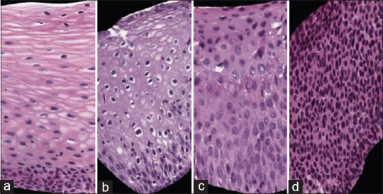
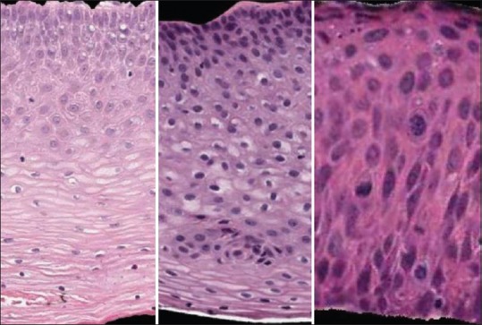
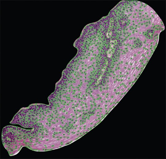
Advances in image analysis and computational techniques have facilitated automatic detection of critical features in histopathology images. Detection of nuclei is critical for squamous epithelium cervical intraepithelial neoplasia (CIN) classification into normal, CIN1, CIN2, and CIN3 grades.

METHODS

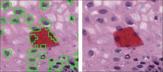
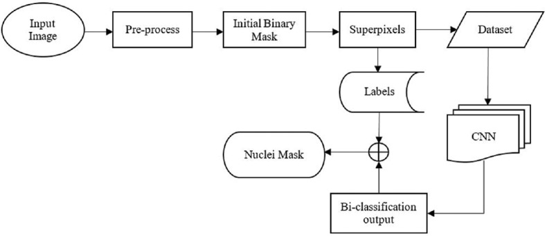
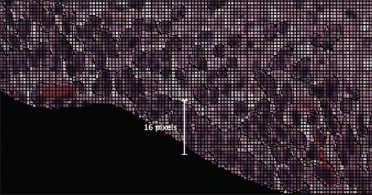
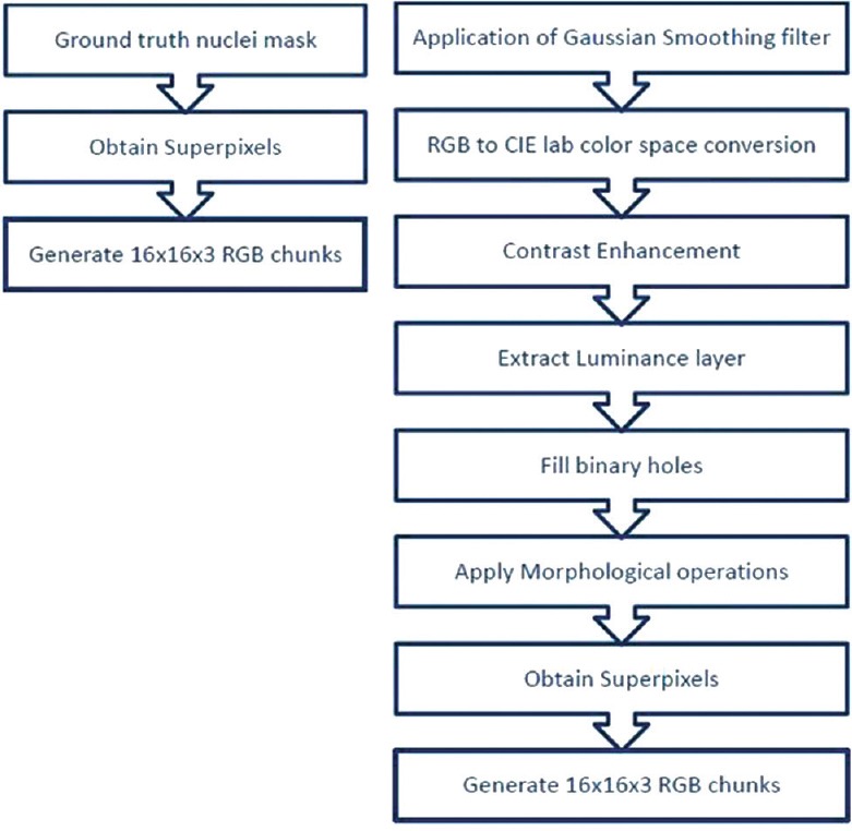
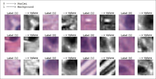
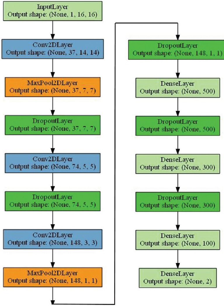
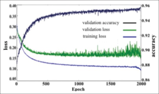
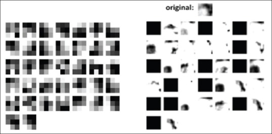
In this study, a deep learning (DL)-based nuclei segmentation approach is investigated based on gathering localized information through the generation of superpixels using a simple linear iterative clustering algorithm and training with a convolutional neural network.

RESULTS

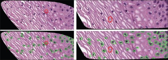
The proposed approach was evaluated on a dataset of 133 digitized histology images and achieved an overall nuclei detection (object-based) accuracy of 95.97%, with demonstrated improvement over imaging-based and clustering-based benchmark techniques.

CONCLUSIONS

The proposed DL-based nuclei segmentation Method with superpixel analysis has shown improved segmentation results in comparison to state-of-the-art methods.

摘要

背景

图像分析和计算技术的进步促进了组织病理学图像中关键特征的自动检测。细胞核的检测对于将鳞状上皮宫颈上皮内瘤变(CIN)分类为正常、CIN1、CIN2和CIN3级至关重要。

方法

在本研究中,基于使用简单线性迭代聚类算法生成超像素来收集局部信息并通过卷积神经网络进行训练,研究了一种基于深度学习(DL)的细胞核分割方法。

结果

该方法在一个包含133张数字化组织学图像的数据集上进行了评估,实现了总体细胞核检测(基于对象)的准确率为95.97%,与基于成像和基于聚类的基准技术相比有明显改进。

结论

与现有技术方法相比,所提出的基于深度学习的带有超像素分析的细胞核分割方法显示出了更好的分割结果。

https://cdn.ncbi.nlm.nih.gov/pmc/blobs/046a/5869967/2da5ee30f149/JPI-9-5-g015.jpg
https://cdn.ncbi.nlm.nih.gov/pmc/blobs/046a/5869967/f0fb0e816260/JPI-9-5-g001.jpg
https://cdn.ncbi.nlm.nih.gov/pmc/blobs/046a/5869967/6ac54a478abd/JPI-9-5-g002.jpg
https://cdn.ncbi.nlm.nih.gov/pmc/blobs/046a/5869967/ca220aec00ea/JPI-9-5-g003.jpg
https://cdn.ncbi.nlm.nih.gov/pmc/blobs/046a/5869967/f6ceb6820286/JPI-9-5-g004.jpg
https://cdn.ncbi.nlm.nih.gov/pmc/blobs/046a/5869967/0335c9eea0a4/JPI-9-5-g005.jpg
https://cdn.ncbi.nlm.nih.gov/pmc/blobs/046a/5869967/cfc1a7461944/JPI-9-5-g006.jpg
https://cdn.ncbi.nlm.nih.gov/pmc/blobs/046a/5869967/495f1e2fa3ba/JPI-9-5-g007.jpg
https://cdn.ncbi.nlm.nih.gov/pmc/blobs/046a/5869967/06579804873f/JPI-9-5-g008.jpg
https://cdn.ncbi.nlm.nih.gov/pmc/blobs/046a/5869967/c2215deec973/JPI-9-5-g009.jpg
https://cdn.ncbi.nlm.nih.gov/pmc/blobs/046a/5869967/9b31223c3b96/JPI-9-5-g010.jpg
https://cdn.ncbi.nlm.nih.gov/pmc/blobs/046a/5869967/2f38c05b11ed/JPI-9-5-g011.jpg
https://cdn.ncbi.nlm.nih.gov/pmc/blobs/046a/5869967/075a7242fec7/JPI-9-5-g013.jpg
https://cdn.ncbi.nlm.nih.gov/pmc/blobs/046a/5869967/2da5ee30f149/JPI-9-5-g015.jpg

相似文献

1
Deep Learning Nuclei Detection in Digitized Histology Images by Superpixels.通过超像素进行数字化组织学图像中的深度学习细胞核检测
J Pathol Inform. 2018 Mar 5;9:5. doi: 10.4103/jpi.jpi_74_17. eCollection 2018.
2
Enhancements in localized classification for uterine cervical cancer digital histology image assessment.子宫颈癌数字组织学图像评估中局部分类的改进。
J Pathol Inform. 2016 Dec 30;7:51. doi: 10.4103/2153-3539.197193. eCollection 2016.
3
EpithNet: Deep Regression for Epithelium Segmentation in Cervical Histology Images.EpithNet:用于宫颈组织学图像上皮细胞分割的深度回归
J Pathol Inform. 2020 Mar 30;11:10. doi: 10.4103/jpi.jpi_53_19. eCollection 2020.
4
Automated Diagnosis of Cervical Intraepithelial Neoplasia in Histology Images via Deep Learning.通过深度学习实现组织学图像中宫颈上皮内瘤变的自动诊断
Diagnostics (Basel). 2022 Feb 21;12(2):548. doi: 10.3390/diagnostics12020548.
5
Nuclei-Based Features for Uterine Cervical Cancer Histology Image Analysis With Fusion-Based Classification.基于细胞核特征的子宫颈癌组织学图像分析及融合分类法
IEEE J Biomed Health Inform. 2016 Nov;20(6):1595-1607. doi: 10.1109/JBHI.2015.2483318. Epub 2015 Oct 26.
6
An automatic nuclei segmentation method based on deep convolutional neural networks for histopathology images.一种基于深度卷积神经网络的组织病理学图像细胞核自动分割方法。
BMC Biomed Eng. 2019 Oct 17;1:24. doi: 10.1186/s42490-019-0026-8. eCollection 2019.
7
DeepCIN: Attention-Based Cervical histology Image Classification with Sequential Feature Modeling for Pathologist-Level Accuracy.深度宫颈影像网络(DeepCIN):基于注意力机制的宫颈组织学图像分类,采用序列特征建模以实现病理学家级别的准确率。
J Pathol Inform. 2020 Dec 24;11:40. doi: 10.4103/jpi.jpi_50_20. eCollection 2020.
8
Automatic cell segmentation in histopathological images via two-staged superpixel-based algorithms.基于两阶段超像素算法的组织病理学图像中自动细胞分割。
Med Biol Eng Comput. 2019 Mar;57(3):653-665. doi: 10.1007/s11517-018-1906-0. Epub 2018 Oct 16.
9
A Bottom-Up Approach for Pancreas Segmentation Using Cascaded Superpixels and (Deep) Image Patch Labeling.一种使用级联超像素和(深度)图像块标记的自下而上的胰腺分割方法。
IEEE Trans Image Process. 2017 Jan;26(1):386-399. doi: 10.1109/TIP.2016.2624198. Epub 2016 Nov 1.
10
Automated Cervical Digitized Histology Whole-Slide Image Analysis Toolbox.自动化宫颈数字化组织学全切片图像分析工具箱
J Pathol Inform. 2021 Jun 9;12:26. doi: 10.4103/jpi.jpi_52_20. eCollection 2021.

引用本文的文献

1
Artificial Intelligence in Medicine: A Specialty-Level Overview of Emerging AI Trends.医学中的人工智能:新兴人工智能趋势的专业层面概述。
JSLS. 2025 Jul-Sep;29(3). doi: 10.4293/JSLS.2025.00041. Epub 2025 Sep 3.
2
Automatic Blob Detection Method for Cancerous Lesions in Unsupervised Breast Histology Images.无监督乳腺组织学图像中癌性病变的自动斑点检测方法
Bioengineering (Basel). 2025 Mar 31;12(4):364. doi: 10.3390/bioengineering12040364.
3
Advancements in pathology: Digital transformation, precision medicine, and beyond.病理学的进展:数字转型、精准医学及其他。

本文引用的文献

1
DeepPap: Deep Convolutional Networks for Cervical Cell Classification.DeepPap:用于宫颈细胞分类的深度卷积神经网络。
IEEE J Biomed Health Inform. 2017 Nov;21(6):1633-1643. doi: 10.1109/JBHI.2017.2705583. Epub 2017 May 19.
2
Automatic Nuclei Detection Based on Generalized Laplacian of Gaussian Filters.基于广义拉普拉斯高斯滤波器的自动细胞核检测。
IEEE J Biomed Health Inform. 2017 May;21(3):826-837. doi: 10.1109/JBHI.2016.2544245. Epub 2016 Mar 21.
3
Deep Voting: A Robust Approach Toward Nucleus Localization in Microscopy Images.
J Pathol Inform. 2024 Nov 19;16:100408. doi: 10.1016/j.jpi.2024.100408. eCollection 2025 Jan.
4
Artificial intelligence across oncology specialties: current applications and emerging tools.肿瘤学各专业中的人工智能:当前应用与新兴工具
BMJ Oncol. 2024 Jan 17;3(1):e000134. doi: 10.1136/bmjonc-2023-000134. eCollection 2024.
5
LAMA: Lesion-Aware Mixup Augmentation for Skin Lesion Segmentation.LAMA:基于病灶感知的图像混合增强在皮肤病灶分割中的应用。
J Imaging Inform Med. 2024 Aug;37(4):1812-1823. doi: 10.1007/s10278-024-01000-5. Epub 2024 Feb 26.
6
Histological classification of canine and feline lymphoma using a modular approach based on deep learning and advanced image processing.基于深度学习和先进图像处理的模块化方法对犬猫淋巴瘤进行组织学分类。
Sci Rep. 2023 Nov 9;13(1):19436. doi: 10.1038/s41598-023-46607-w.
7
Efficient Convolution Network to Assist Breast Cancer Diagnosis and Target Therapy.用于辅助乳腺癌诊断和靶向治疗的高效卷积网络。
Cancers (Basel). 2023 Aug 6;15(15):3991. doi: 10.3390/cancers15153991.
8
Artificial Intelligence in pathology: current applications, limitations, and future directions.人工智能在病理学中的应用:现状、局限和未来方向。
Ir J Med Sci. 2024 Apr;193(2):1117-1121. doi: 10.1007/s11845-023-03479-3. Epub 2023 Aug 5.
9
Refining skin lesions classification performance using geometric features of superpixels.利用超像素的几何特征改进皮肤损伤分类性能。
Sci Rep. 2023 Jul 15;13(1):11463. doi: 10.1038/s41598-023-38706-5.
10
Skin Lesion Segmentation in Dermoscopic Images with Noisy Data.带噪声数据的皮肤镜图像皮肤损伤分割。
J Digit Imaging. 2023 Aug;36(4):1712-1722. doi: 10.1007/s10278-023-00819-8. Epub 2023 Apr 5.
深度投票:一种用于显微镜图像中细胞核定位的稳健方法。
Med Image Comput Comput Assist Interv. 2015 Oct;9351:374-382. doi: 10.1007/978-3-319-24574-4_45. Epub 2015 Nov 18.
4
Multi-Pass Adaptive Voting for Nuclei Detection in Histopathological Images.多遍自适应投票法在病理图像细胞核检测中的应用。
Sci Rep. 2016 Oct 3;6:33985. doi: 10.1038/srep33985.
5
When machine vision meets histology: A comparative evaluation of model architecture for classification of histology sections.当机器视觉遇上组织学:用于组织学切片分类的模型架构比较评估
Med Image Anal. 2017 Jan;35:530-543. doi: 10.1016/j.media.2016.08.010. Epub 2016 Sep 9.
6
Robust Nucleus/Cell Detection and Segmentation in Digital Pathology and Microscopy Images: A Comprehensive Review.在数字病理学和显微镜图像中进行稳健的细胞核/细胞检测和分割:全面综述。
IEEE Rev Biomed Eng. 2016;9:234-63. doi: 10.1109/RBME.2016.2515127. Epub 2016 Jan 6.
7
Nuclei-Based Features for Uterine Cervical Cancer Histology Image Analysis With Fusion-Based Classification.基于细胞核特征的子宫颈癌组织学图像分析及融合分类法
IEEE J Biomed Health Inform. 2016 Nov;20(6):1595-1607. doi: 10.1109/JBHI.2015.2483318. Epub 2015 Oct 26.
8
An Automatic Learning-Based Framework for Robust Nucleus Segmentation.一种基于自动学习的稳健细胞核分割框架。
IEEE Trans Med Imaging. 2016 Feb;35(2):550-66. doi: 10.1109/TMI.2015.2481436. Epub 2015 Sep 23.
9
Stacked Sparse Autoencoder (SSAE) for Nuclei Detection on Breast Cancer Histopathology Images.用于乳腺癌组织病理学图像细胞核检测的堆叠稀疏自动编码器(SSAE)
IEEE Trans Med Imaging. 2016 Jan;35(1):119-30. doi: 10.1109/TMI.2015.2458702. Epub 2015 Jul 20.
10
Mitosis detection in breast cancer pathology images by combining handcrafted and convolutional neural network features.通过结合手工制作的特征和卷积神经网络特征来检测乳腺癌病理图像中的有丝分裂。
J Med Imaging (Bellingham). 2014 Oct;1(3):034003. doi: 10.1117/1.JMI.1.3.034003. Epub 2014 Oct 10.