Domínguez-Vicent Alberto, Monsálvez-Romín Daniel, Esteve-Taboada José J, Montés-Micó Robert, Ferrer-Blasco Teresa
Department of Optics and Optometry and Vision Sciences, University of Valencia, Spain.
Department of Optics and Optometry and Vision Sciences, University of Valencia, Spain.
J Optom. 2019 Jan-Mar;12(1):14-21. doi: 10.1016/j.optom.2018.01.001. Epub 2018 Apr 4.
To compare changes in the ciliary muscle area at different sectors between pre-presbyopic and presbyopic eyes during accommodation by means of an anterior segment optical coherence tomographer (OCT).
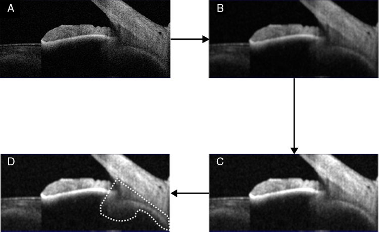
The anterior ciliary muscle area was measured in 20 healthy and phakic pre-presbyopic eyes, whose mean age was 23.3±4.4 years, and in 20 healthy and phakic presbyopic eyes, whose mean age was 46.5±5.2 years. The relative change in the cross-sectional area of the ciliary muscle was measured at the nasal, inferior, and temporal sectors between 0 and -3 D of vergence, in -1 D step. A linear model was used to assess the correlation of each eye parameter with the accommodative demand.
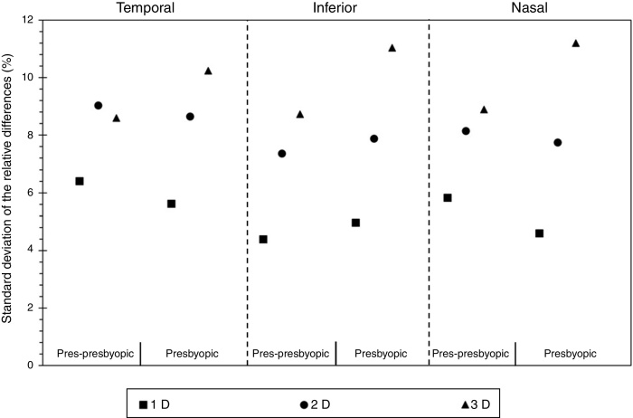
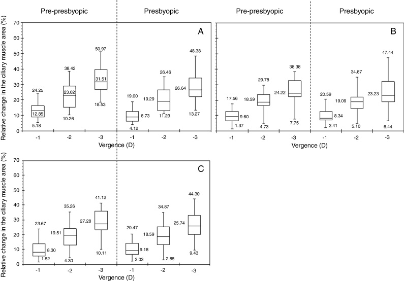
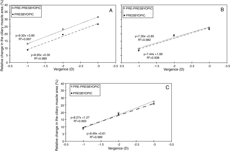
Each population group showed a significant increase in the anterior ciliary muscle area for each sector. The maximum increase in the ciliary muscle area within the pre-presbyopic group was about 30%, and for the presbyopic one was about 25%. At the same time, it was obtained that the larger the vergence, the larger the variability. Furthermore, the linear model showed a positive tendency between the change in the ciliary muscle area of each sector and the vergence for both population groups, which coefficient of determination was in all cases greater than 0.93.
The anterior ciliary muscle area tends to increase with accommodation. The presbyopic nasal, inferior, and temporal ciliary muscle seem to have the same contractile capability as the young presbyopic ciliary muscle. These results might help to increase the evidences in the knowledge regarding the modern understanding of accommodation biometry and biomechanics.
通过前段光学相干断层扫描仪(OCT)比较老视前期和老视眼在调节过程中不同象限睫状肌面积的变化。
测量20只健康有晶状体老视前期眼(平均年龄23.3±4.4岁)和20只健康有晶状体老视眼(平均年龄46.5±5.2岁)的睫状肌前面积。在0至-3D的聚散度之间,以-1D步长测量鼻侧、下方和颞侧象限睫状肌横截面积的相对变化。使用线性模型评估每个眼参数与调节需求的相关性。
每组人群各象限的睫状肌前面积均显著增加。老视前期组睫状肌面积的最大增加约为30%,老视组约为25%。同时发现,聚散度越大,变异性越大。此外,线性模型显示两组人群各象限睫状肌面积变化与聚散度之间呈正相关趋势,所有情况下决定系数均大于0.93。
睫状肌前面积倾向于随调节增加。老视眼的鼻侧、下方和颞侧睫状肌似乎与年轻老视前期睫状肌具有相同的收缩能力。这些结果可能有助于增加关于调节生物测量学和生物力学现代理解的知识证据。