Mohamed Alizae Marny, Wong Kiong Hung, Lee Wan Jen, Marizan Nor Murshida, Mohd Hussaini Haizal, Rosli Tanti Irawati
Department of Orthodontics, Faculty of Dentistry, Universiti Kebangsaan Malaysia, Kuala Lumpur, Malaysia.
Ministry of Health, Malaysia.
Saudi Dent J. 2018 Apr;30(2):142-150. doi: 10.1016/j.sdentj.2017.12.001. Epub 2018 Jan 4.
The aim of the study was to evaluate the effect of resin infiltration on colour changes and surface roughness of artificial white spot lesions (WSLs) on maxillary and mandibular premolar.
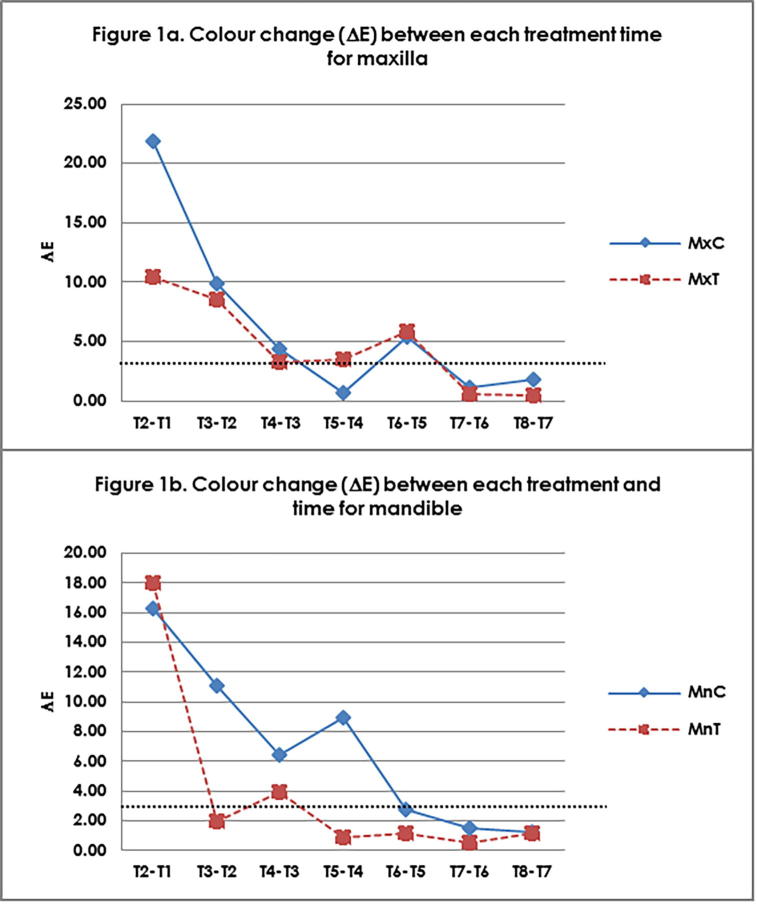
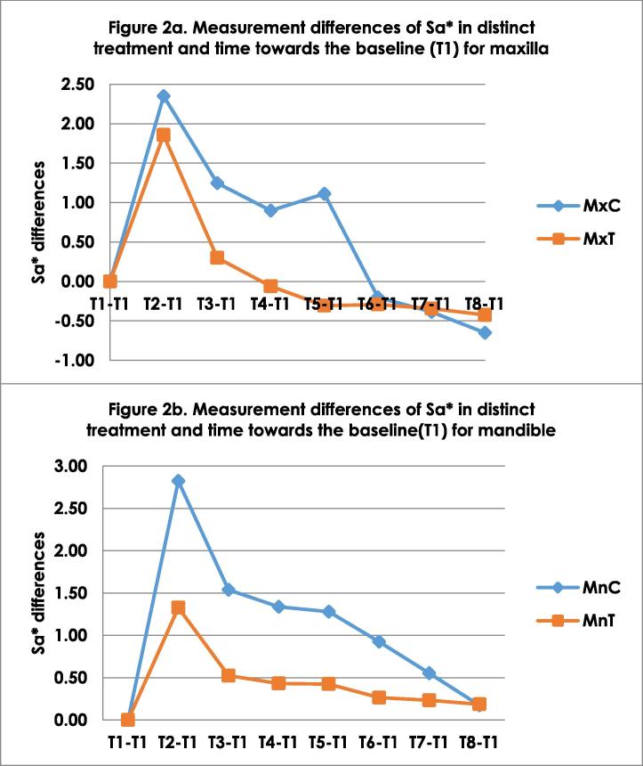
Sixty (60) extracted sound Maxilla (Mx) and Mandibular (Mn) premolars were randomly divided into 2 groups (test and control). Artificial WSLs were produced on buccal surface of teeth and were immersed in artificial saliva for 8 weeks. Colour components (L, a, b) and surface roughness (Sa) were assessed on 40 teeth using colour difference meter RD-100 and Alicona® Infinite Focus profilometer respectively. The measurements were done at baseline (T1), directly after artificial WSLs (T2), after 24 hours immersed in saliva and application of resin (T3) and immersion in artificial saliva for 1 (T4), 2 (T5), 4 (T6), 6 (T7) and 8 (T8) weeks. SEM images analysis were carried out on 20 teeth in four time points.
The values of L (lightness), b (yellow/blue) and Sa (surface roughness) are gradually reduced to the baseline value. Whereas, the value of a gradually increased with distinct treatment time to achieve the baseline value. The higher value of L and Sa, the whiter the lesion suggesting higher degree of enamel demineralization and surface roughness. Lower L values suggest a masking colour effect.
The material produced favorable esthetics on colour and the surface roughness of teeth at distinct treatment times. It is recommended to be used to improve WSL post orthodontic treatment.
本研究旨在评估树脂渗透对上颌和下颌前磨牙人工白斑病变(WSLs)颜色变化和表面粗糙度的影响。
60颗拔除的完好上颌(Mx)和下颌(Mn)前磨牙随机分为2组(试验组和对照组)。在牙齿颊面制作人工WSLs,并将其浸泡在人工唾液中8周。分别使用色差仪RD - 100和Alicona® Infinite Focus轮廓仪对40颗牙齿的颜色成分(L、a、b)和表面粗糙度(Sa)进行评估。测量在基线(T1)、人工WSLs制作后即刻(T2)、浸泡在唾液中并应用树脂24小时后(T3)以及浸泡在人工唾液中1(T4)、2(T5)、4(T6)、6(T7)和8(T8)周时进行。在四个时间点对20颗牙齿进行扫描电子显微镜(SEM)图像分析。
L(亮度)、b(黄/蓝)和Sa(表面粗糙度)的值逐渐降低至基线值。而a值随着不同的治疗时间逐渐增加以达到基线值。L和Sa值越高,病变越白,表明釉质脱矿程度和表面粗糙度越高。较低的L值表明有掩盖颜色的效果。
该材料在不同治疗时间对牙齿的颜色和表面粗糙度产生了良好的美学效果。建议用于正畸治疗后改善WSLs。