Xu Xiaojun, Guan Xiaojun, Guo Tao, Zeng Qiaoling, Ye Rong, Wang Jiaqiu, Zhong Jianguo, Xuan Min, Gu Quanquan, Huang Peiyu, Pu Jiali, Zhang Baorong, Zhang Minming
Department of Radiology, Second Affiliated Hospital of Zhejiang University School of Medicine, Hangzhou, China.
Department of Neurology, Second Affiliated Hospital of Zhejiang University School of Medicine, Hangzhou, China.
Front Hum Neurosci. 2018 Mar 27;12:117. doi: 10.3389/fnhum.2018.00117. eCollection 2018.
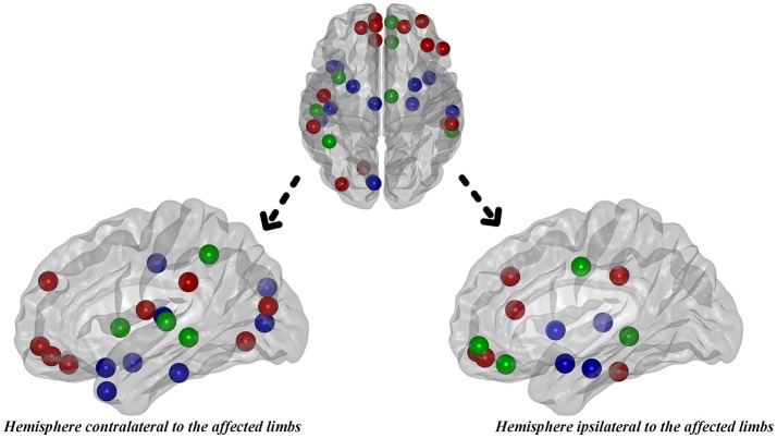
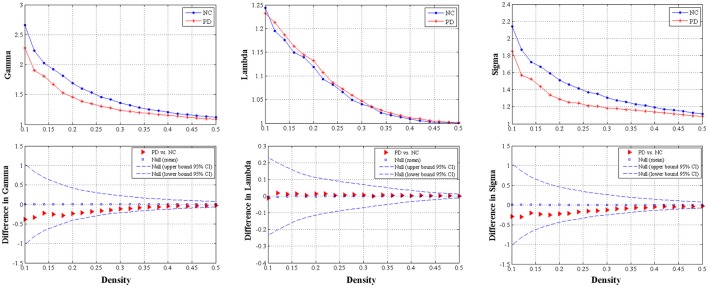
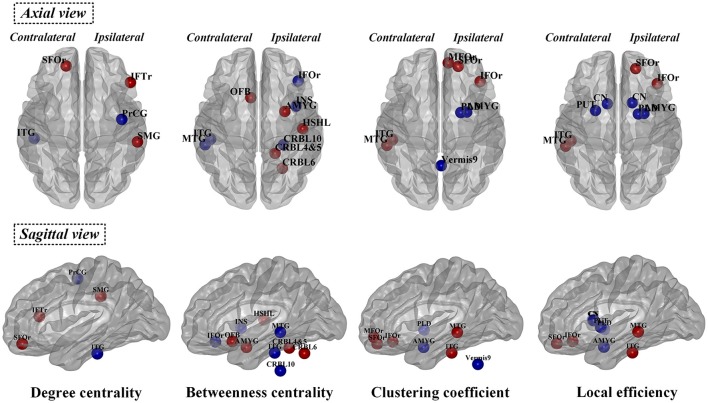
Hemiparkinsonism duration in patients with Parkinson's disease (PD) is a key time window to study early pathology of PD. We aimed to comprehensively explore the alterations of deformation and structural network in PD patients with hemiparkinsonism, which could potentially disclose the early biomarker for PD. Thirty-one PD patients with hemiparkinsonism and 37 age- and gender- matched normal controls were included in the present study. First of all, we normalized the left hemisphere of structural images as the contralateral side to the affected limbs. Deformation-based morphometry (DBM) was conducted to evaluate the brain atrophy and/or enlargement. structural networks were constructed by thresholding gray matter volume correlation matrices of 116 regions and analyzed using graph theoretical approaches (e.g., small-worldness, global, and nodal measures). Significantly decreased deformation values were observed in the temporoparietal regions like bilateral middle temporal gyri, ipsilateral precuneus and contralateral Rolandic operculum extending to supramarginal and postcentral gyri. Lower deformation values in contralateral middle temporal gyrus were negatively correlated with higher motor impairment which was dominated by akinesia/rigidity. Moreover, nodal reorganization of structural network mainly located in frontal, temporal, subcortex and cerebellum was bilaterally explored in PD patients with hemiparkinsonism. Increased nodal properties could be commonly observed in frontal lobes. Disruption of subcortex including basal ganglia and amygdala was detected by nodal local efficiency and nodal clustering coefficient. Twelve hubs, mainly from paralimbic-limbic and heteromodal networks, were disrupted and, alternatively, 14 hubs, most of which were located in frontal lobes, were additionally detected in PD patients with hemiparkinsonism. In conclusion, during hemiparkinsonism period, mild brain atrophy in the temporoparietal regions and widespread reorganization of structural network, e.g., enhanced frontal function and disruption of basal ganglia nodes, occurred in both hemispheres. With our data, we can also argue that MTG contralateral to the affected limbs (expressing clinically verified brain atrophy) might be a potential living biomarker to monitor disease progression. Therefore, the combination of DBM and structural network analyses can provide a comprehensive and sensitive evaluation for potential pathogenesis of early PD patients with hemiparkinsonism.
帕金森病(PD)患者的偏侧帕金森病持续时间是研究PD早期病理的关键时间窗口。我们旨在全面探索偏侧帕金森病PD患者的形变和结构网络改变,这可能揭示PD的早期生物标志物。本研究纳入了31例偏侧帕金森病患者和37例年龄及性别匹配的正常对照。首先,我们将结构图像的左半球归一化为患侧肢体对侧的半球。采用基于形变的形态学测量(DBM)来评估脑萎缩和/或脑扩大。通过对116个区域的灰质体积相关矩阵进行阈值处理构建结构网络,并使用图论方法(如小世界特性、全局和节点测量)进行分析。在颞顶叶区域观察到显著降低的形变值,如双侧颞中回、同侧楔前叶和对侧中央沟盖延伸至缘上回和中央后回。对侧颞中回较低的形变值与以运动不能/强直为主的较高运动障碍呈负相关。此外,在偏侧帕金森病PD患者中双侧探索了主要位于额叶、颞叶、皮层下和小脑的结构网络节点重组。在额叶通常可观察到节点属性增加。通过节点局部效率和节点聚类系数检测到包括基底神经节和杏仁核在内的皮层下破坏。在偏侧帕金森病PD患者中,12个主要来自边缘旁 - 边缘和异模态网络的枢纽被破坏,另外还检测到14个枢纽,其中大部分位于额叶。总之,在偏侧帕金森病期间,颞顶叶区域出现轻度脑萎缩,结构网络广泛重组,例如双侧额叶功能增强和基底神经节节点破坏。基于我们的数据,我们还可以认为患侧肢体对侧的颞中回(表现出经临床验证的脑萎缩)可能是监测疾病进展的潜在活体生物标志物。因此,DBM和结构网络分析的结合可以为偏侧帕金森病早期患者的潜在发病机制提供全面而敏感的评估。