Hoekel James, Narayanan Anagha, Rutlin Jerrel, Lugar Heather, Al-Lozi Amal, Hershey Tamara, Tychsen Lawrence
Department of Ophthalmology, Washington University School of Medicine and St. Louis Children's Hospital, St. Louis, Missouri, USA.
Department of Psychiatry, Washington University School of Medicine, St. Louis, Missouri, USA.
BMJ Open Ophthalmol. 2018 Jan 18;3(1):e000081. doi: 10.1136/bmjophth-2017-000081. eCollection 2018.
BACKGROUND/AIMS: To report alterations in visual acuity and visual pathway structure over an interval of 1-3 years in a cohort of children, adolescents and young adults who have Wolfram syndrome (WFS) and to describe the range of disease severity evident in patients with WFS whose ages differed by as much as 20 years at first examination.
Annual, prospective ophthalmological examinations were performed in conjunction with retinal nerve fibre layer (RNFL) analysis. Diffusion tensor MRI-derived fractional anisotropy was used to assess the microstructural integrity of the optic radiations (OR FA).
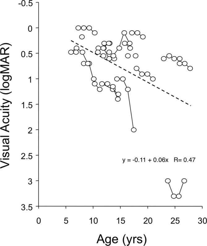
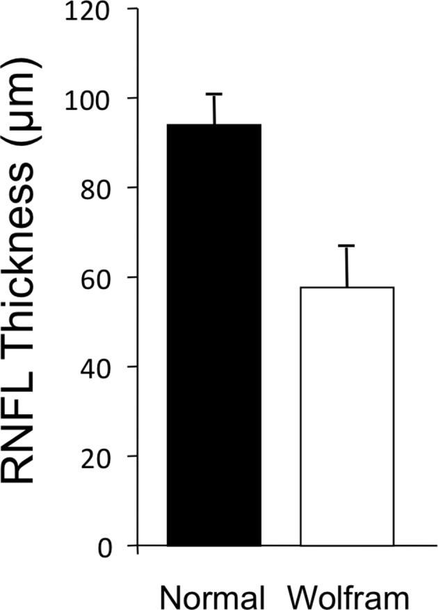
Mean age of the 23 patients with WFS in the study was 13.8 years (range 5-25 years). Mean log minimum angle resolution visual acuity was 0.66 (20/91). RNFL thickness was subnormal in even the youngest patients with WFS. Average RNFL thickness in patients with WFS was 57±8 µ or ~40% thinner than that measured in normal (94±10 µ) children and adolescents (P<0.01). Lower OR FA correlated with worse visual acuity (P=0.006). Subsequent examinations showed declines (P<0.05) in visual acuity, RNFL thickness and OR FA at follow-up intervals of 12-36 months. However, a wide range of disease severity was evident across ages: some of the youngest patients at their first examination had deficits more severe than the oldest patients.
The genetic mutation of WFS causes damage to both pregeniculate and postgeniculate regions of the visual pathway. The damage is progressive. The decline in visual pathway structure is accompanied by declines of visual function. Disease severity differs widely in individual patients and cannot be predicted from their age.
背景/目的:报告患有沃夫勒姆综合征(WFS)的儿童、青少年和年轻成年人在1至3年期间视力和视觉通路结构的变化,并描述初次检查时年龄相差多达20岁的WFS患者中明显的疾病严重程度范围。
每年进行前瞻性眼科检查,并结合视网膜神经纤维层(RNFL)分析。使用扩散张量MRI衍生的分数各向异性来评估视辐射的微观结构完整性(OR FA)。
该研究中23例WFS患者的平均年龄为13.8岁(范围5 - 25岁)。平均对数最小分辨角视力为0.66(20/91)。即使是最年轻的WFS患者,RNFL厚度也低于正常水平。WFS患者的平均RNFL厚度为57±8µ,比正常儿童和青少年(94±10µ)测量值薄约40%(P<0.01)。较低的OR FA与较差的视力相关(P = 0.006)。后续检查显示,在12 - 36个月的随访期间,视力、RNFL厚度和OR FA均下降(P<0.05)。然而,各年龄段的疾病严重程度差异很大:一些初次检查时最年轻的患者存在比最年长患者更严重的缺陷。
WFS的基因突变会对视神经通路的膝状体前和膝状体后区域造成损害。这种损害是渐进性的。视觉通路结构的下降伴随着视觉功能的下降。个体患者的疾病严重程度差异很大,无法根据其年龄预测。