Kovacs-Kàsa Anita, Varn Matthew N, Verin Alexander D, Gonzales Joyce N
Vascular Biology Center, Augusta University Health, Augusta, USA.
Division of Pulmonary and Critical Care Medicine, Department of Internal Medicine, Augusta University Health, Augusta, USA.
Sci Pages Pulmonol. 2017;1(1):7-18.
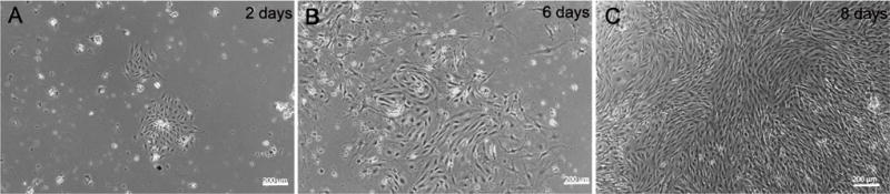
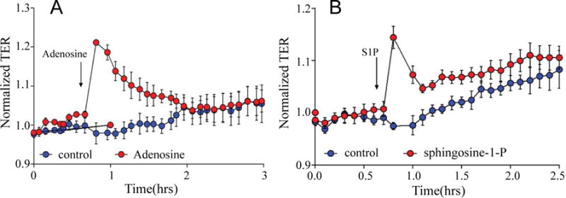
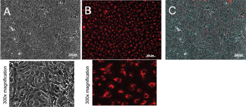
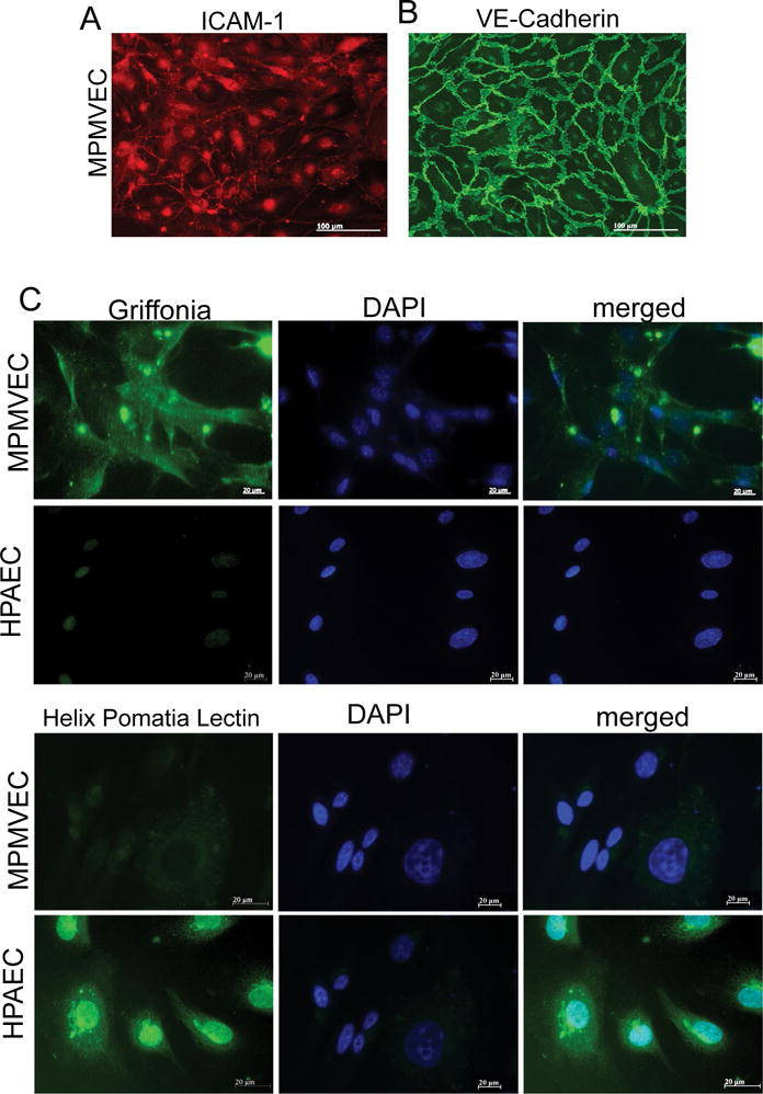
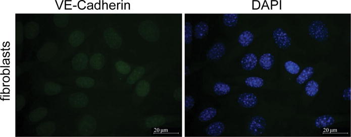
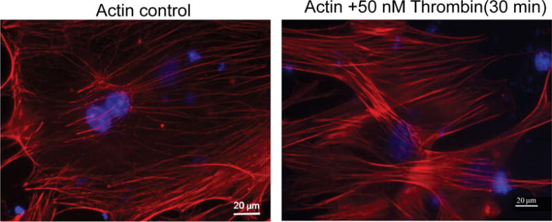
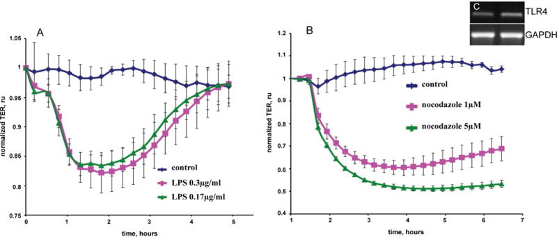
Pulmonary microvascular endothelial cells (ECs) are integral to the alveoli-capillary barrier of the lung. The EC barrier integrity is known to be disrupted in severe lung diseases such as acute respiratory distress syndrome (ARDS), pneumonia and pulmonary edema. Mice are commonly used to model these diseases, dictating an increasingly high demand for murine ECs isolation and culture. Despite the significant number of protocols for the culture of various types of murine cells, the isolation of microvascular endothelial cells remains a challenging procedure. In our manuscript we developed adetailed step-by-step refined method for isolation murine pulmonary microvascular ECs for studies. We separated cells using platelet endothelial cell adhesion molecule antibody and characterized ECs with antibodies against intercellular adhesion molecule-1, acetylated-low density lipoprotein, and vascular endothelial (VE)-cadherin. Further, we confirmed microvascular origin of these cells using (negative control) staining. Barrier properties of EC monolayer were characterized by conducting electric cell-substrate impedance sensing experiments with the edemagenic agents, lipopolysaccharide and nocodazole, and known barrier-protective agents, adenosine and sphingosine-1-phosphate. The described complete protocol provided consistent and reproducible results.
肺微血管内皮细胞(ECs)是肺肺泡-毛细血管屏障的重要组成部分。已知在严重肺部疾病如急性呼吸窘迫综合征(ARDS)、肺炎和肺水肿中,EC屏障的完整性会受到破坏。小鼠常用于模拟这些疾病,这使得对小鼠ECs分离和培养的需求日益增加。尽管有大量关于各种类型小鼠细胞培养的方案,但微血管内皮细胞的分离仍然是一个具有挑战性的过程。在我们的手稿中,我们开发了一种详细的逐步改进方法,用于分离小鼠肺微血管ECs以进行研究。我们使用血小板内皮细胞黏附分子抗体分离细胞,并用抗细胞间黏附分子-1、乙酰化低密度脂蛋白和血管内皮(VE)-钙黏蛋白的抗体对ECs进行表征。此外,我们使用(阴性对照)染色确认了这些细胞的微血管起源。通过用电细胞-基质阻抗传感实验,使用致水肿剂脂多糖和诺考达唑以及已知的屏障保护剂腺苷和鞘氨醇-1-磷酸,对EC单层的屏障特性进行了表征。所描述的完整方案提供了一致且可重复的结果。