• 文献检索
  • 文档翻译
  • 深度研究
  • 学术资讯
  • Suppr Zotero 插件Zotero 插件
  • 邀请有礼
  • 套餐&价格
  • 历史记录
应用&插件
Suppr Zotero 插件Zotero 插件浏览器插件Mac 客户端Windows 客户端微信小程序
定价
高级版会员购买积分包购买API积分包
服务
文献检索文档翻译深度研究API 文档MCP 服务
关于我们
关于 Suppr公司介绍联系我们用户协议隐私条款
关注我们

Suppr 超能文献

核心技术专利:CN118964589B侵权必究
粤ICP备2023148730 号-1Suppr @ 2026

文献检索

告别复杂PubMed语法,用中文像聊天一样搜索,搜遍4000万医学文献。AI智能推荐,让科研检索更轻松。

立即免费搜索

文件翻译

保留排版,准确专业,支持PDF/Word/PPT等文件格式,支持 12+语言互译。

免费翻译文档

深度研究

AI帮你快速写综述,25分钟生成高质量综述,智能提取关键信息,辅助科研写作。

立即免费体验

血管分泌鞘氨醇 1-磷酸通过激活肺泡 II 型细胞中的 S1PR2-YAP 信号轴对于肺修复至关重要。

Angiocrine Sphingosine-1-Phosphate Activation of S1PR2-YAP Signaling Axis in Alveolar Type II Cells Is Essential for Lung Repair.

机构信息

Department of Pharmacology and Regenerative Medicine, University of Illinois College of Medicine, Chicago, IL 60612, USA.

Department of Bioengineering, University of Illinois at Chicago, Chicago, IL 60607, USA.

出版信息

Cell Rep. 2020 Jun 30;31(13):107828. doi: 10.1016/j.celrep.2020.107828.

DOI:10.1016/j.celrep.2020.107828
PMID:32610129
原文链接:https://pmc.ncbi.nlm.nih.gov/articles/PMC7371431/
Abstract

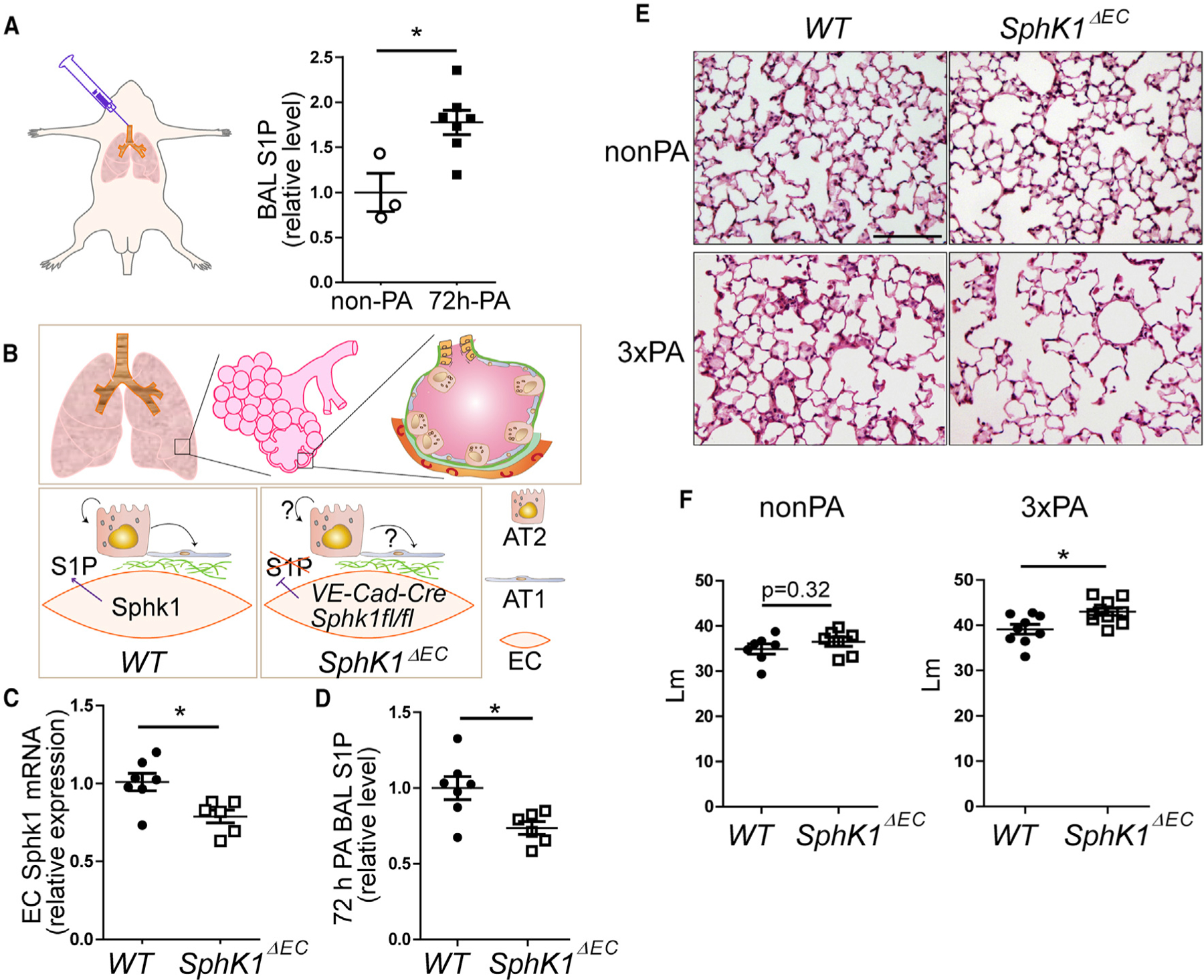
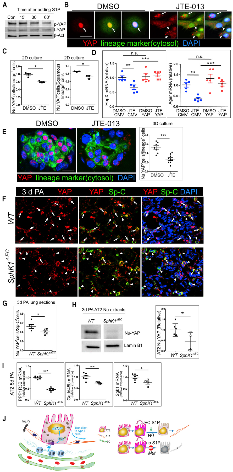
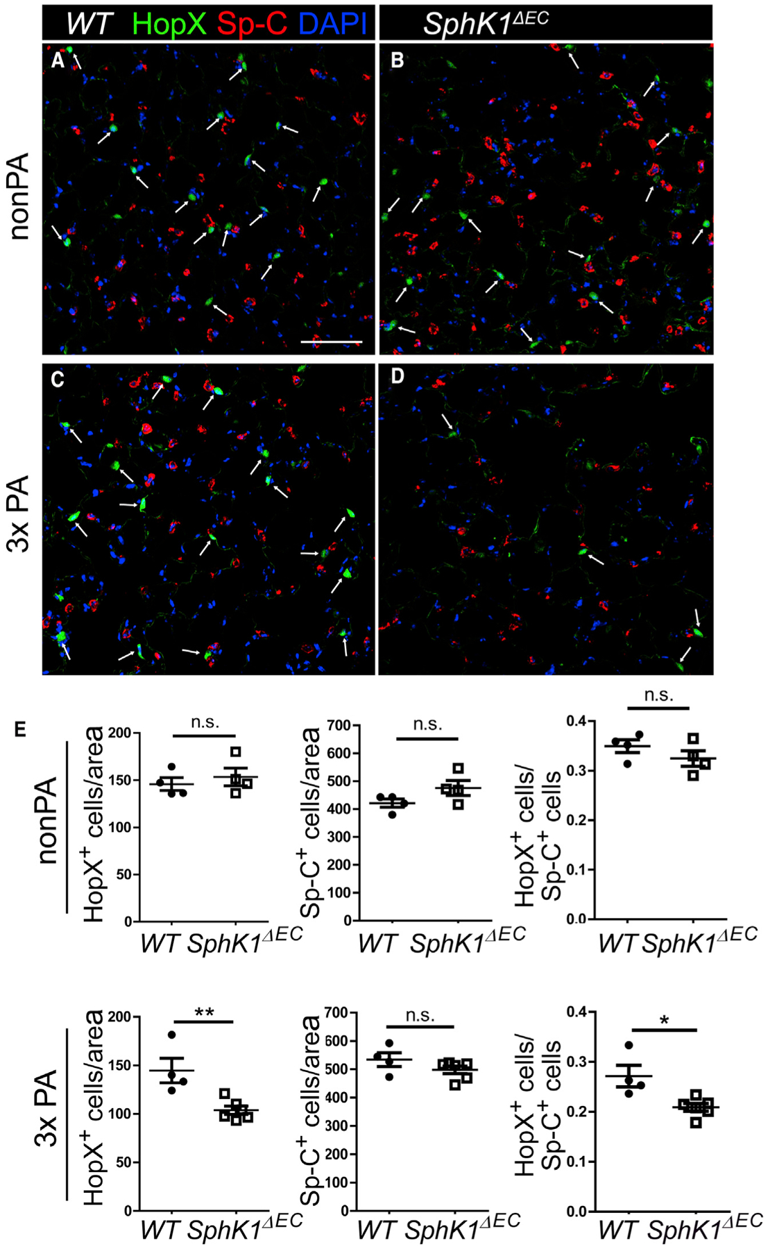
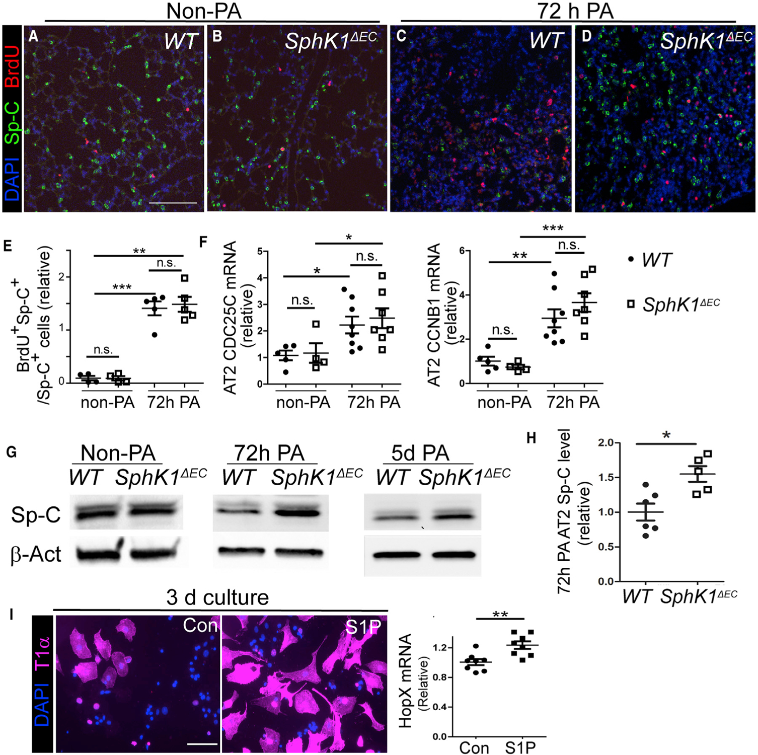
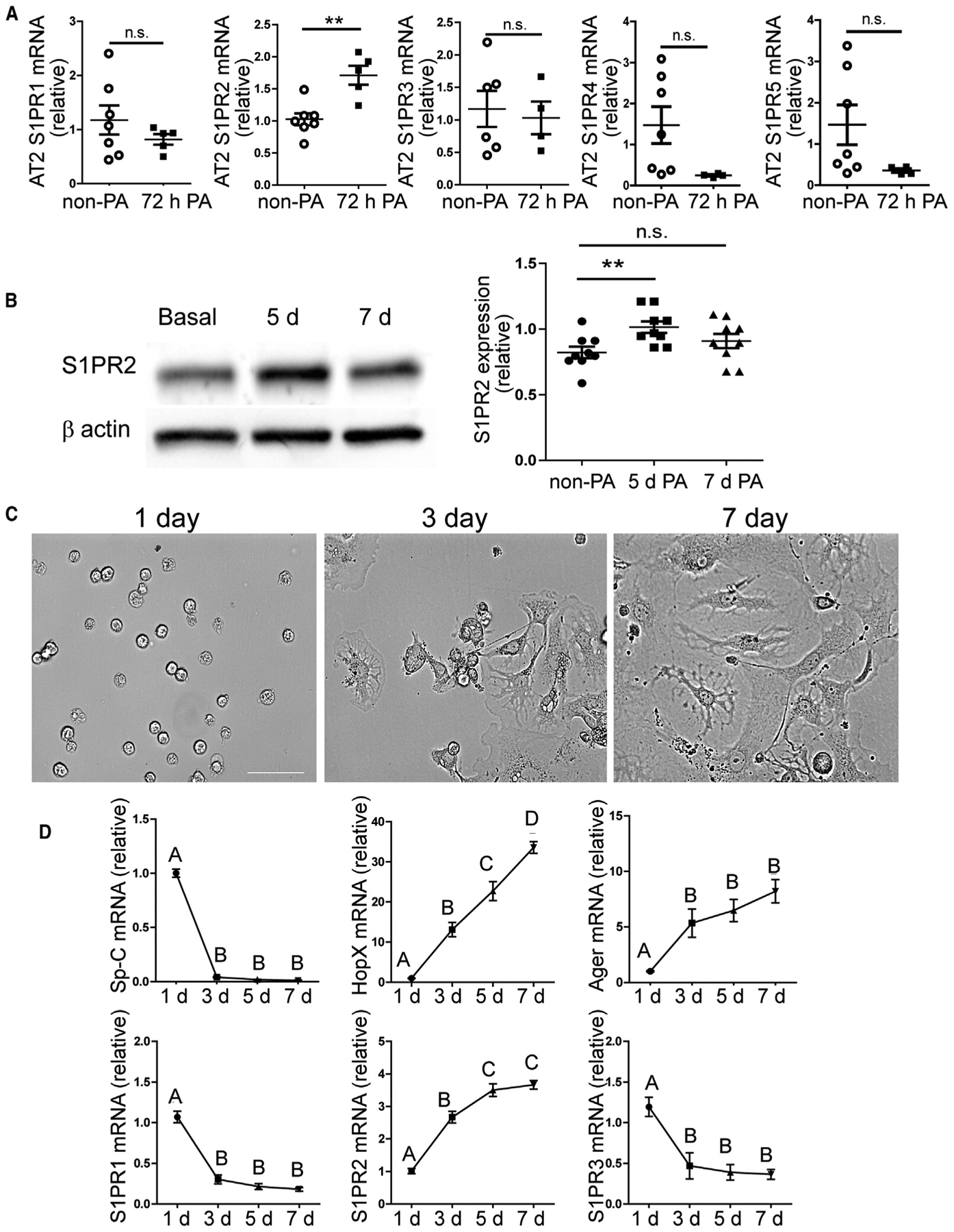
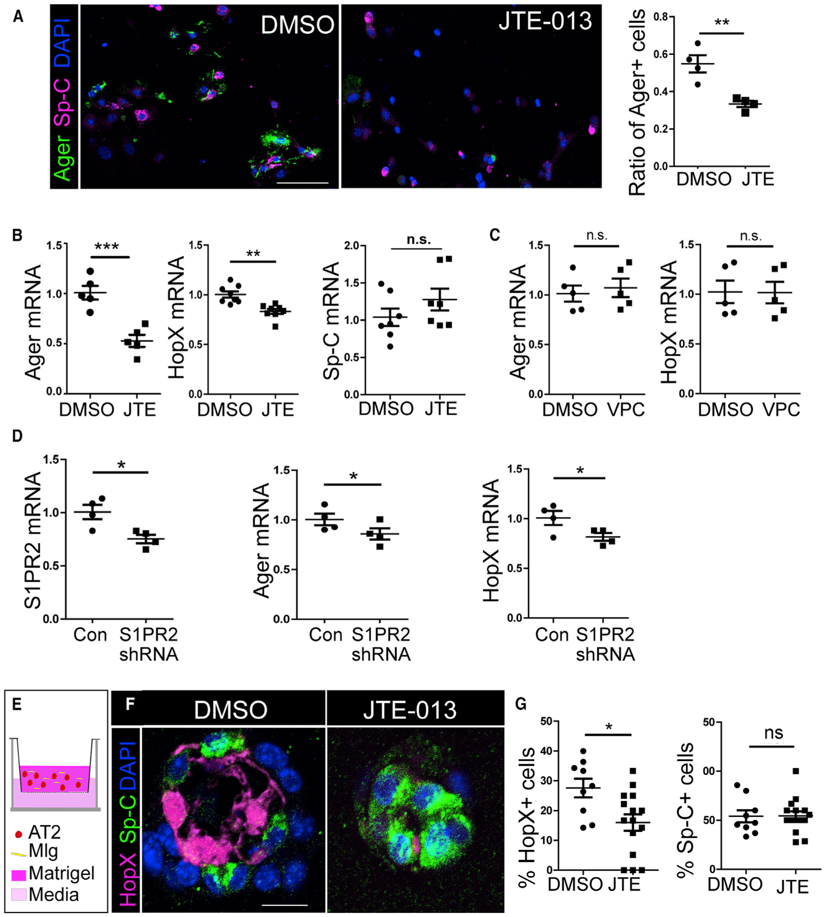
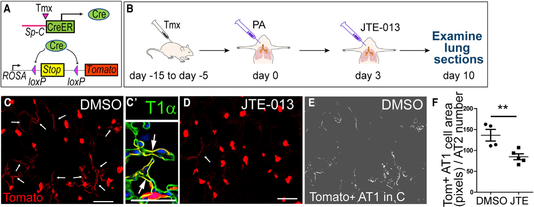
Lung alveolar epithelium is composed of alveolar type I (AT1) and type II (AT2) cells. AT1 cells mediate gas exchange, whereas AT2 cells act as progenitor cells to repair injured alveoli. Lung microvascular endothelial cells (LMVECs) play a crucial but still poorly understood role in regulating alveolar repair. Here, we studied the role of the LMVEC-derived bioactive lipid sphingosine-1-phosphate (S1P) in promoting alveolar repair using mice with endothelial-specific deletion of sphingosine kinase 1 (Sphk1), the key enzyme promoting S1P generation. These mutant lungs developed airspace-enlargement lesions and exhibited a reduced number of AT1 cells after Pseudomonas-aeruginosa-induced lung injury. We demonstrated that S1P released by LMVECs acted via its receptor, S1PR2, on AT2 cells and induced nuclear translocation of yes-associated protein (YAP), a regulator of AT2 to AT1 transition. Thus, angiocrine S1P released after injury acts via the S1PR2-YAP signaling axis on AT2 cells to promote AT2 to AT1 differentiation required for alveolar repair.

摘要

肺肺泡上皮由肺泡 I 型 (AT1) 和 II 型 (AT2) 细胞组成。AT1 细胞介导气体交换,而 AT2 细胞作为祖细胞来修复受损的肺泡。肺微血管内皮细胞 (LMVEC) 在调节肺泡修复中起着至关重要但仍知之甚少的作用。在这里,我们使用内皮细胞特异性敲除鞘氨醇激酶 1 (Sphk1) 的小鼠研究了 LMVEC 衍生的生物活性脂质 1-磷酸鞘氨醇 (S1P) 在促进肺泡修复中的作用,Sphk1 是促进 S1P 生成的关键酶。这些突变肺在铜绿假单胞菌诱导的肺损伤后发展出气腔扩大病变,并表现出 AT1 细胞数量减少。我们证明,LMVEC 释放的 S1P 通过其受体 S1PR2 作用于 AT2 细胞,并诱导 yes 相关蛋白 (YAP) 的核易位,YAP 是 AT2 向 AT1 转化的调节因子。因此,损伤后释放的血管旁 S1P 通过 S1PR2-YAP 信号轴作用于 AT2 细胞,促进肺泡修复所需的 AT2 向 AT1 分化。

https://cdn.ncbi.nlm.nih.gov/pmc/blobs/b050/7371431/a0d23464211e/nihms-1608489-f0008.jpg
https://cdn.ncbi.nlm.nih.gov/pmc/blobs/b050/7371431/2cfd1bd9b40b/nihms-1608489-f0002.jpg
https://cdn.ncbi.nlm.nih.gov/pmc/blobs/b050/7371431/1308b2cca9ec/nihms-1608489-f0003.jpg
https://cdn.ncbi.nlm.nih.gov/pmc/blobs/b050/7371431/afb8538e8845/nihms-1608489-f0004.jpg
https://cdn.ncbi.nlm.nih.gov/pmc/blobs/b050/7371431/55b129a01429/nihms-1608489-f0005.jpg
https://cdn.ncbi.nlm.nih.gov/pmc/blobs/b050/7371431/e3678b6ad7bb/nihms-1608489-f0006.jpg
https://cdn.ncbi.nlm.nih.gov/pmc/blobs/b050/7371431/6dd45529b633/nihms-1608489-f0007.jpg
https://cdn.ncbi.nlm.nih.gov/pmc/blobs/b050/7371431/a0d23464211e/nihms-1608489-f0008.jpg

相似文献

1
Angiocrine Sphingosine-1-Phosphate Activation of S1PR2-YAP Signaling Axis in Alveolar Type II Cells Is Essential for Lung Repair.血管分泌鞘氨醇 1-磷酸通过激活肺泡 II 型细胞中的 S1PR2-YAP 信号轴对于肺修复至关重要。
Cell Rep. 2020 Jun 30;31(13):107828. doi: 10.1016/j.celrep.2020.107828.
2
Sphingosine Kinase 1/S1P Signaling Contributes to Pulmonary Fibrosis by Activating Hippo/YAP Pathway and Mitochondrial Reactive Oxygen Species in Lung Fibroblasts.鞘氨醇激酶 1/S1P 信号通过激活肺成纤维细胞中的 Hippo/YAP 通路和线粒体活性氧物质促进肺纤维化。
Int J Mol Sci. 2020 Mar 17;21(6):2064. doi: 10.3390/ijms21062064.
3
Pancreas lineage allocation and specification are regulated by sphingosine-1-phosphate signalling.胰腺谱系分配和特化受1-磷酸鞘氨醇信号调控。
PLoS Biol. 2017 Mar 1;15(3):e2000949. doi: 10.1371/journal.pbio.2000949. eCollection 2017 Mar.
4
Activation of sphingosine kinase-1 reverses the increase in lung vascular permeability through sphingosine-1-phosphate receptor signaling in endothelial cells.鞘氨醇激酶-1的激活通过内皮细胞中1-磷酸鞘氨醇受体信号传导逆转肺血管通透性的增加。
Circ Res. 2008 Nov 7;103(10):1164-72. doi: 10.1161/01.RES.0000338501.84810.51. Epub 2008 Oct 10.
5
Epidermal growth factor-induced cellular invasion requires sphingosine-1-phosphate/sphingosine-1-phosphate 2 receptor-mediated ezrin activation.表皮生长因子诱导的细胞侵袭需要鞘氨醇-1-磷酸/鞘氨醇-1-磷酸 2 受体介导的 ezrin 激活。
FASEB J. 2013 Aug;27(8):3155-66. doi: 10.1096/fj.13-228460. Epub 2013 Apr 29.
6
The sphingosine-1-phosphate/sphingosine-1-phosphate receptor 2 axis regulates early airway T-cell infiltration in murine mast cell-dependent acute allergic responses.1-磷酸鞘氨醇/1-磷酸鞘氨醇受体2轴在小鼠肥大细胞依赖性急性过敏反应中调节早期气道T细胞浸润。
J Allergy Clin Immunol. 2015 Apr;135(4):1008-1018.e1. doi: 10.1016/j.jaci.2014.10.044. Epub 2014 Dec 13.
7
Sphingosine-1-phosphate enhances satellite cell activation in dystrophic muscles through a S1PR2/STAT3 signaling pathway.鞘氨醇-1-磷酸通过 S1PR2/STAT3 信号通路增强萎缩肌肉中的卫星细胞激活。
PLoS One. 2012;7(5):e37218. doi: 10.1371/journal.pone.0037218. Epub 2012 May 14.
8
Exogenous S1P via S1P receptor 2 induces CTGF expression through Src-RhoA-ROCK-YAP pathway in hepatic stellate cells.外源性 S1P 通过 S1P 受体 2 通过Src-RhoA-ROCK-YAP 通路诱导肝星状细胞中 CTGF 的表达。
Mol Biol Rep. 2024 Sep 2;51(1):950. doi: 10.1007/s11033-024-09868-w.
9
Sphingosine kinase 1 deficiency confers protection against hyperoxia-induced bronchopulmonary dysplasia in a murine model: role of S1P signaling and Nox proteins.鞘氨醇激酶 1 缺乏可保护小鼠免受高氧诱导的支气管肺发育不良:S1P 信号和 Nox 蛋白的作用。
Am J Pathol. 2013 Oct;183(4):1169-1182. doi: 10.1016/j.ajpath.2013.06.018. Epub 2013 Aug 8.
10
Role of sphingosine-1-phosphate (S1P) and the S1P(2) receptor in allergen-induced, mast cell-dependent contraction of rat lung parenchymal strips.鞘氨醇-1-磷酸(S1P)及S1P(2)受体在变应原诱导的、肥大细胞依赖性大鼠肺实质条收缩中的作用
Naunyn Schmiedebergs Arch Pharmacol. 2009 Oct;380(4):303-9. doi: 10.1007/s00210-009-0438-4. Epub 2009 Jul 28.

引用本文的文献

1
New perspectives on the progression of pulmonary fibrosis: the cascade from aberrant microvascular endothelial cell activation to fibrosis.肺纤维化进展的新视角:从异常微血管内皮细胞激活到纤维化的级联反应。
Front Med (Lausanne). 2025 Aug 21;12:1639043. doi: 10.3389/fmed.2025.1639043. eCollection 2025.
2
Conjugated bile acids promote metabolic dysfunction-associated steatotic liver disease through inducing nuclear translocation of sphingosine-1-phosphate receptor 2 to disrupt peroxisome proliferator-activated receptor alpha.结合胆汁酸通过诱导鞘氨醇-1-磷酸受体2的核转位以破坏过氧化物酶体增殖物激活受体α,从而促进代谢功能障碍相关脂肪性肝病。
Cell Commun Signal. 2025 May 25;23(1):240. doi: 10.1186/s12964-025-02249-1.
3

本文引用的文献

1
Defining the role of pulmonary endothelial cell heterogeneity in the response to acute lung injury.定义肺血管内皮细胞异质性在急性肺损伤反应中的作用。
Elife. 2020 Feb 24;9:e53072. doi: 10.7554/eLife.53072.
2
Epithelial Vegfa Specifies a Distinct Endothelial Population in the Mouse Lung.上皮细胞 Vegfa 在小鼠肺中特异性指定了一个独特的血管内皮细胞群体。
Dev Cell. 2020 Mar 9;52(5):617-630.e6. doi: 10.1016/j.devcel.2020.01.009. Epub 2020 Feb 13.
3
Yap/Taz regulate alveolar regeneration and resolution of lung inflammation.Yap/Taz 调节肺泡再生和肺炎症的解决。
High matrix stiffness promotes senescence of type II alveolar epithelial cells by lysosomal degradation of lamin A/C in pulmonary fibrosis.
高基质硬度通过肺纤维化中核纤层蛋白A/C的溶酶体降解促进II型肺泡上皮细胞衰老。
Respir Res. 2025 Apr 9;26(1):128. doi: 10.1186/s12931-025-03201-0.
4
Blood flow-induced angiocrine signals promote organ growth and regeneration.血流诱导的血管分泌信号促进器官生长和再生。
Bioessays. 2025 Feb;47(2):e2400207. doi: 10.1002/bies.202400207. Epub 2024 Nov 11.
5
Unleashing AdipoRon's Potential: A Fresh Approach to Tackle Infections in Bronchiectasis via Sphingosine Metabolism Modulation.释放脂联素受体激动剂(AdipoRon)的潜力:通过鞘氨醇代谢调节应对支气管扩张症感染的新方法。
J Inflamm Res. 2024 Oct 24;17:7653-7674. doi: 10.2147/JIR.S483689. eCollection 2024.
6
Long-term pulmonary repair in rat lungs after sublobar resection: electrocautery versus stapler methods.大鼠肺叶下切除术后的长期肺修复:电灼法与吻合器法对比
Gen Thorac Cardiovasc Surg. 2024 Oct 28. doi: 10.1007/s11748-024-02098-8.
7
Pulmonary fibrosis: pathogenesis and therapeutic strategies.肺纤维化:发病机制与治疗策略。
MedComm (2020). 2024 Sep 23;5(10):e744. doi: 10.1002/mco2.744. eCollection 2024 Oct.
8
YAP1 inhibits the senescence of alveolar epithelial cells by targeting Prdx3 to alleviate pulmonary fibrosis.YAP1 通过靶向 Prdx3 抑制肺泡上皮细胞衰老,从而减轻肺纤维化。
Exp Mol Med. 2024 Jul;56(7):1643-1654. doi: 10.1038/s12276-024-01277-0. Epub 2024 Jul 1.
9
Novel insights into the potential applications of stem cells in pulmonary hypertension therapy.干细胞在肺动脉高压治疗中潜在应用的新见解。
Respir Res. 2024 Jun 7;25(1):237. doi: 10.1186/s12931-024-02865-4.
10
Characterization of perivascular alveolar epithelial stem cells and their niche in lung homeostasis and cancer.肺稳态和癌症中血管周围肺泡上皮干细胞及其龛的特征。
Stem Cell Reports. 2024 Jun 11;19(6):890-905. doi: 10.1016/j.stemcr.2024.04.009. Epub 2024 May 16.
J Clin Invest. 2019 Apr 15;129(5):2107-2122. doi: 10.1172/JCI125014.
4
Single cell RNA sequencing identifies TGFβ as a key regenerative cue following LPS-induced lung injury.单细胞 RNA 测序鉴定 TGFβ 为 LPS 诱导的肺损伤后关键的再生信号。
JCI Insight. 2019 Mar 26;5(8):123637. doi: 10.1172/jci.insight.123637.
5
Dlk1-Mediated Temporal Regulation of Notch Signaling Is Required for Differentiation of Alveolar Type II to Type I Cells during Repair.Dlk1 介导的 Notch 信号时空调节对于修复过程中肺泡 II 型细胞向 I 型细胞的分化是必需的。
Cell Rep. 2019 Mar 12;26(11):2942-2954.e5. doi: 10.1016/j.celrep.2019.02.046.
6
stimulates nuclear sphingosine-1-phosphate generation and epigenetic regulation of lung inflammatory injury.刺激核鞘氨醇-1-磷酸的产生和肺炎症损伤的表观遗传调控。
Thorax. 2019 Jun;74(6):579-591. doi: 10.1136/thoraxjnl-2018-212378. Epub 2019 Feb 5.
7
inactivation reveals Hippo function in alveolar type I cell differentiation during lung transition to air breathing.失活揭示 Hippo 在肺向空气呼吸过渡过程中肺泡 I 型细胞分化中的作用。
Development. 2018 Nov 9;145(21):dev163105. doi: 10.1242/dev.163105.
8
The Hippo Signaling Network and Its Biological Functions.Hippo 信号通路及其生物学功能。
Annu Rev Genet. 2018 Nov 23;52:65-87. doi: 10.1146/annurev-genet-120417-031621. Epub 2018 Sep 5.
9
Method for the Culture of Mouse Pulmonary Microvascular Endothelial Cells.小鼠肺微血管内皮细胞的培养方法
Sci Pages Pulmonol. 2017;1(1):7-18.
10
Flow Cytometry Underestimates and Planimetry Overestimates Alveolar Epithelial Type 2 Cell Expansion after Lung Injury.流式细胞术低估而平面测量法高估肺损伤后肺泡Ⅱ型上皮细胞的增殖。
Am J Respir Crit Care Med. 2018 Aug 1;198(3):390-392. doi: 10.1164/rccm.201709-1838LE.