Tong Ying, Yin Yong, Cheng Pinjing, Lu Jie, Liu Tonghai, Chen Jinhu, Gong Guanzhong
Radiation Physics Department of Shandong Cancer Hospital (Affiliated to Shandong University), Road Jiyan, No. 440, Jinan, Shandong, China.
School of Nuclear Science and Technology, University of South China, Hengyang, China.
J Radiat Res. 2018 Jul 1;59(4):462-468. doi: 10.1093/jrr/rry026.
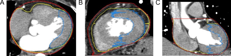
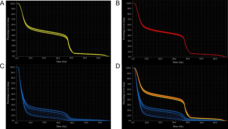
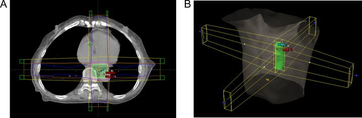
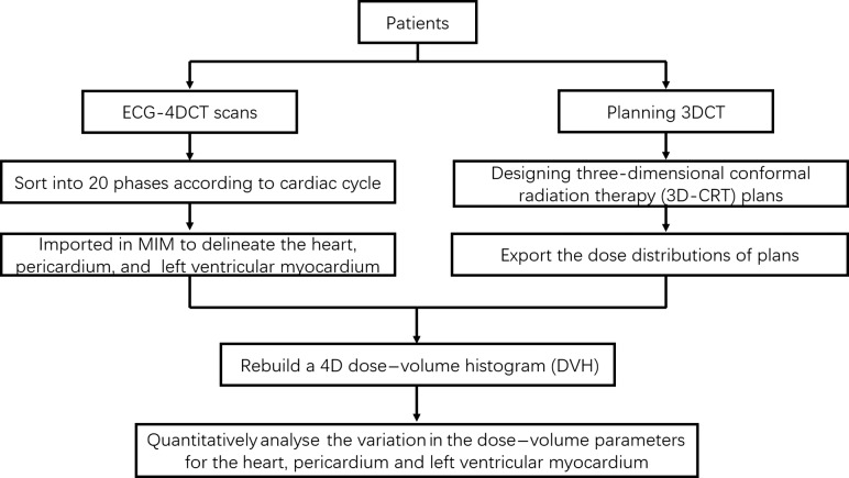
Cardiac activity can induce dose-volume evaluation errors for cardiac structures. The purpose of this study was to quantify the variation in dose-volume parameters for the heart, pericardium and left ventricular myocardium (LVM) throughout the cardiac circle. The heart, pericardium and LVM of 22 patients were contoured on 20 phases of electrocardiography-gated 4D computed tomography (4DCT) images acquired during breath-hold. Radiotherapy plans were designed on 0% phase of the 4DCT images, and the dose distributions of the plans were imported into MIM Maestro and deformed to each phase to generate distributions for all phases. Variations in dose-volume parameters for the heart, pericardium and LVM were compared among different phases. The rates of variation in Dmean for the heart and pericardium were 3.33 ± 1.04% and 2.66 ± 1.15%, respectively. The mean values of the maximum difference in V5, V10, V20, V30 and V40 were all <2% for the heart and pericardium and were not statistically significant (P > 0.05). The rate of variation in Dmean for the LVM reached 87.05 ± 38.34%, and the maximum differences in V5, V10, V20, V30 and V40 were 13.76 ± 4.46%, 13.64 ± 4.33%, 12.84 ± 4.55%, 11.62 ± 4.85% and 3.63 ± 2.56%, respectively; all differences were statistically significant (P < 0.05). Variations in dose-volume parameters were more significant in the LVM than in the heart and pericardium (P < 0.05). The dose-volume parameters for the LVM were significantly influenced by cardiac activity, whereas those for the heart and pericardium were not; therefore, individual dosimetric evaluation and limitation must be performed for the LVM.
心脏活动可导致心脏结构的剂量体积评估误差。本研究的目的是量化整个心动周期中心脏、心包和左心室心肌(LVM)的剂量体积参数变化。在屏气期间采集的22例患者的心电图门控4D计算机断层扫描(4DCT)图像的20个时相上对心脏、心包和LVM进行轮廓勾画。在4DCT图像的0%时相设计放射治疗计划,并将计划的剂量分布导入MIM Maestro并变形至每个时相,以生成所有时相的分布。比较不同时相中心脏、心包和LVM的剂量体积参数变化。心脏和心包的平均剂量(Dmean)变化率分别为3.33±1.04%和2.66±1.15%。心脏和心包的V5、V10、V20、V30和V40的最大差异平均值均<2%,且无统计学意义(P>0.05)。LVM的Dmean变化率达到87.05±38.34%,V5、V10、V20、V30和V40的最大差异分别为13.76±4.46%、13.64±4.33%、12.84±4.55%、11.62±4.85%和3.63±2.56%;所有差异均有统计学意义(P<0.05)。LVM的剂量体积参数变化比心脏和心包更显著(P<0.05)。LVM的剂量体积参数受心脏活动的显著影响,而心脏和心包的则不受影响;因此,必须对LVM进行个体剂量学评估和限制。