Radiation Physics Department of Shandong Cancer Hospital Affiliated to Shandong University, Jinan, China.
School of Nuclear Science and Technology, University of South China, Hengyang, China.
Radiat Oncol. 2018 Aug 10;13(1):145. doi: 10.1186/s13014-018-1093-z.
The deformable image registration (DIR) technique has the potential to realize the dose accumulation during radiotherapy. This study will analyze the feasibility of evaluating dose-volume parameters for the heart and left ventricular myocardium (LVM) by applying DIR.
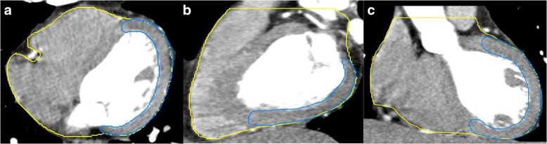
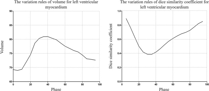
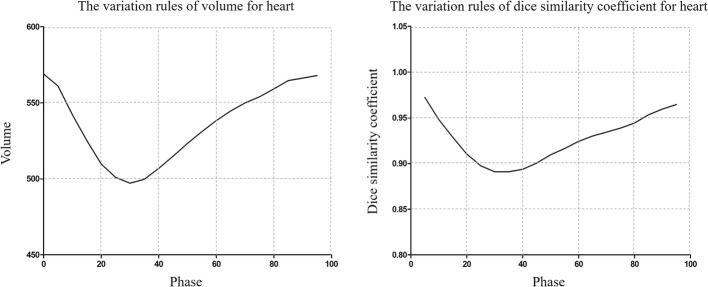
The electrocardiograph-gated four-dimensional CT (ECG-gated 4DCT) data of 21 patients were analyzed retrospectively. The heart and LVM were contoured on 20 phases of 4DCT (0%, 5%,…,95%). The heart and LVM in the minimum volume/dice similarity coefficient (DSC) phase (Volume /DSC ) were deformed to the maximum volume/DSC phase (Volume / DSC ), which used the intensity-based free-form DIR algorithm of MIM software. The dose was deformed according to the deformation vector. The variations in volume, mean dose (D), V, V and V for the heart and LVM before and after DIR were compared, and the reference phase was the Volume /DSC phase.
For the heart, the difference between the pre- and post-registration Volume and Volume were reduced from 13.87 to 1.72%; the DSC was increased from 0.899 to 0.950 between the pre- and post-registration DSC phase relative to the DSC phase. The post-registration D, V, V and V of the heart were statistically significant compared to those in the Volume /DSC phase (p < 0.05). For the LVM, the difference between the pre- and post-registration Volume and Volume were only reduced from 18.77 to 17.38%; the DSC reached only 0.733 in the post-registration DSC phase relative to the DSC phase. The pre- and post-registration volume, D, V, V and V of the LVM were all statistically significant compared to those in the Volume /DSC phase (p < 0.05).
There was no significant relationship between the variation in dose-volume parameters and the variation in the volume and morphology for the heart; however, the inconsistency of the variation in the volume and morphology for the LVM was a major factor that led to uncertainty in the dose-volume evaluation. In addition, the individualized local deformation registration technology should be applied in dose accumulation for the heart and LVM.
形变图像配准(DIR)技术有望实现放射治疗过程中的剂量积累。本研究将分析应用 DIR 评估心脏和左心室心肌(LVM)剂量-体积参数的可行性。
回顾性分析 21 例患者的心电图门控四维 CT(ECG-gated 4DCT)数据。在 4DCT 的 20 个时相(0%、5%、...、95%)上对心脏和 LVM 进行勾画。将心脏和 LVM 在最小体积/散度相似系数(DSC)时相(Volume /DSC )上的形变到最大体积/DSC 时相(Volume / DSC ),采用 MIM 软件的基于强度的自由形态 DIR 算法。根据变形向量对剂量进行变形。比较心脏和 LVM 形变前后的体积、平均剂量(D)、V、V 和 V 的变化,参考时相为 Volume /DSC 时相。
对于心脏,注册前后的 Volume 和 Volume 之间的差异从 13.87 减少到 1.72%;与 Volume /DSC 时相相比,注册前后的 DSC 从 0.899 增加到 0.950。与 Volume /DSC 时相相比,心脏的注册后 D、V、V 和 V 均具有统计学意义(p<0.05)。对于 LVM,注册前后的 Volume 和 Volume 之间的差异仅从 18.77 减少到 17.38%;与 Volume /DSC 时相相比,注册后的 DSC 仅达到 0.733。与 Volume /DSC 时相相比,LVM 的注册前后的体积、D、V、V 和 V 均具有统计学意义(p<0.05)。
心脏的剂量-体积参数变化与体积和形态变化之间没有显著关系;然而,LVM 体积和形态变化的不一致是导致剂量-体积评估不确定的主要因素。此外,应在心脏和 LVM 的剂量积累中应用个体化局部变形配准技术。