Liu Taoyan, Huang Chengwu, Li Hongxia, Wu Fujian, Luo Jianwen, Lu Wenjing, Lan Feng
Beijing Laboratory for Cardiovascular Precision Medicine, The Key Laboratory of Remodeling-Related Cardiovascular Disease, Ministry of Education, Beijing Collaborative Innovation Center for Cardiovascular Disorders, Anzhen Hospital, Capital Medical University, Beijing 100029, China.
Beijing Institute of Heart, Lung and Blood Vessel Diseases, Beijing 100029, China.
Aging (Albany NY). 2018 Apr 14;10(4):532-548. doi: 10.18632/aging.101411.
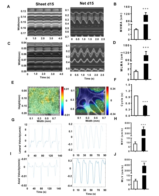
The use of human-induced pluripotent stem cell-derived cardiomyocytes (hiPSC-CMs) is limited in drug discovery and cardiac disease mechanism studies due to cell immaturity. Although many approaches have been reported to improve the maturation of hiPSC-CMs, the elucidation of the process of maturation is crucial. We applied a small-molecule-based differentiation method to generate cardiomyocytes (CMs) with multiple aggregation forms. The motion analysis revealed significant physical differences in the differently shaped CMs, and the net-shaped CMs had larger motion amplitudes and faster velocities than the sheet-shaped CMs. The net-shaped CMs displayed accelerated maturation at the transcriptional level and were more similar to CMs with a prolonged culture time (30 days) than to sheet-d15. Ion channel genes and gap junction proteins were up-regulated in net-shaped CMs, indicating that robust contraction was coupled with enhanced ion channel and connexin expression. The net-shaped CMs also displayed improved myofibril ultrastructure under transmission electron microscopy. In conclusion, different multicellular hPSC-CM structures, such as the net-shaped pattern, are formed using the conditioned induction method, providing a useful tool to improve cardiac maturation.
由于细胞不成熟,人诱导多能干细胞衍生的心肌细胞(hiPSC-CMs)在药物发现和心脏疾病机制研究中的应用受到限制。尽管已有许多方法报道可改善hiPSC-CMs的成熟度,但阐明成熟过程至关重要。我们应用基于小分子的分化方法来生成具有多种聚集形式的心肌细胞(CMs)。运动分析显示不同形状的CMs存在显著的物理差异,网状CMs比片状CMs具有更大的运动幅度和更快的速度。网状CMs在转录水平上显示出加速成熟,并且与培养时间延长(30天)的CMs相比,比片状d15的CMs更相似。离子通道基因和缝隙连接蛋白在网状CMs中上调,表明强烈的收缩与增强的离子通道和连接蛋白表达相关。在透射电子显微镜下,网状CMs的肌原纤维超微结构也得到改善。总之,使用条件诱导方法可形成不同的多细胞hPSC-CM结构,如网状模式,为改善心脏成熟提供了有用的工具。