Laboratory of Parasitic Diseases, National Institute of Allergy and Infectious Diseases, National Institutes of Health, Bethesda, MD, United States of America.
EMD Serono Research and Development Institute, Billerica, MA, United States of America.
PLoS Negl Trop Dis. 2018 Apr 18;12(4):e0006404. doi: 10.1371/journal.pntd.0006404. eCollection 2018 Apr.
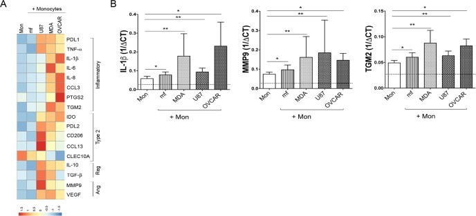
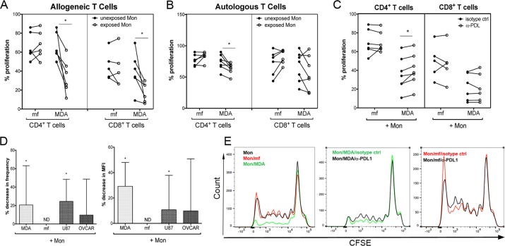
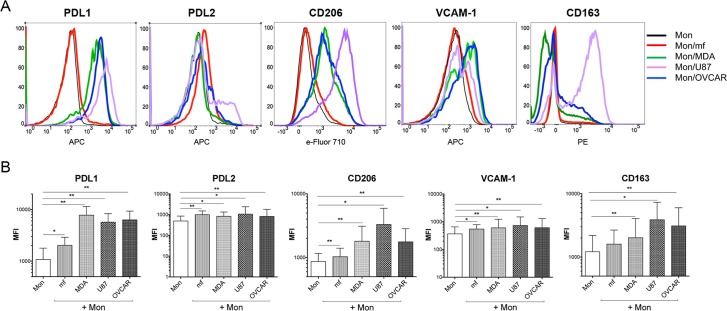
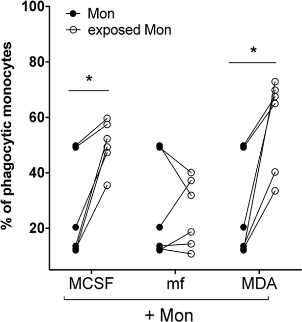
A number of features at the host-parasite interface are reminiscent of those that are also observed at the host-tumor interface. Both cancer cells and parasites establish a tissue microenvironment that allows for immune evasion and may reflect functional alterations of various innate cells. Here, we investigated how the phenotype and function of human monocytes is altered by exposure to cancer cell lines and if these functional and phenotypic alterations parallel those induced by exposure to helminth parasites. Thus, human monocytes were exposed to three different cancer cell lines (breast, ovarian, or glioblastoma) or to live microfilariae (mf) of Brugia malayi-a causative agent of lymphatic filariasis. After 2 days of co-culture, monocytes exposed to cancer cell lines showed markedly upregulated expression of M1-associated (TNF-α, IL-1β), M2-associated (CCL13, CD206), Mreg-associated (IL-10, TGF-β), and angiogenesis associated (MMP9, VEGF) genes. Similar to cancer cell lines, but less dramatically, mf altered the mRNA expression of IL-1β, CCL13, TGM2 and MMP9. When surface expression of the inhibitory ligands PDL1 and PDL2 was assessed, monocytes exposed to both cancer cell lines and to live mf significantly upregulated PDL1 and PDL2 expression. In contrast to exposure to mf, exposure to cancer cell lines increased the phagocytic ability of monocytes and reduced their ability to induce T cell proliferation and to expand Granzyme A+ CD8+ T cells. Our data suggest that despite the fact that helminth parasites and cancer cell lines are extraordinarily disparate, they share the ability to alter the phenotype of human monocytes.
宿主-寄生虫界面的许多特征让人联想到宿主-肿瘤界面上观察到的特征。癌细胞和寄生虫都建立了一个组织微环境,使免疫逃逸成为可能,并可能反映出各种固有细胞的功能改变。在这里,我们研究了暴露于癌细胞系如何改变人单核细胞的表型和功能,以及这些功能和表型的改变是否与暴露于寄生虫引起的改变平行。因此,人单核细胞分别暴露于三种不同的癌细胞系(乳腺癌、卵巢癌或神经胶质瘤)或活的班氏吴策线虫微丝蚴(mf)——淋巴丝虫病的病原体。在共培养 2 天后,暴露于癌细胞系的单核细胞表现出显著上调的 M1 相关(TNF-α,IL-1β)、M2 相关(CCL13,CD206)、Mreg 相关(IL-10,TGF-β)和血管生成相关(MMP9,VEGF)基因的表达。与癌细胞系相似,但程度较轻,mf 改变了 IL-1β、CCL13、TGM2 和 MMP9 的 mRNA 表达。当评估抑制性配体 PDL1 和 PDL2 的表面表达时,暴露于两种癌细胞系和活 mf 的单核细胞显著上调了 PDL1 和 PDL2 的表达。与暴露于 mf 相反,暴露于癌细胞系增加了单核细胞的吞噬能力,降低了其诱导 T 细胞增殖和扩增 Granzyme A+CD8+T 细胞的能力。我们的数据表明,尽管寄生虫和癌细胞系非常不同,但它们都有能力改变人单核细胞的表型。