Department of Cardiology, Sun Yat-sen Memorial Hospital of Sun Yat-sen University, No.107, Yanjiang West Road, Yuexiu District, Guangzhou, 510120, China.
Department of Cardiology, The First Affiliated Hospital of NanChang University, Nanchang, 330006, China.
J Transl Med. 2018 Apr 18;16(1):105. doi: 10.1186/s12967-018-1481-z.
Perivascular adipose tissue (PVAT) accelerates plaque progression and increases cardiovascular risk. We tested the hypothesis that PVAT contributed to plaque vulnerability and investigated whether endoplasmic reticulum stress (ER stress) in PVAT played an important role in vulnerable plaque.
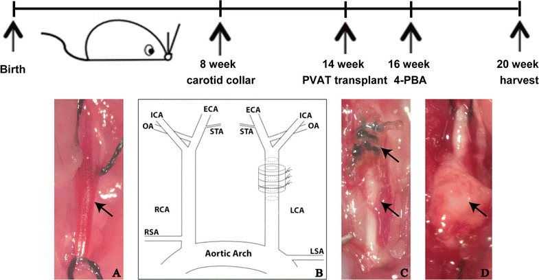
We transplanted thoracic aortic PVAT or subcutaneous adipose tissue as a control, from donor mice to carotid arteries of recipient apolipoprotein E deficient (apoE) mice after removing carotid artery collar placed for 6 weeks. Two weeks after transplantation, ER stress inhibitor 4-phenyl butyric acid (4-PBA) was locally administrated to the transplanted PVAT and then animals were euthanized after 4 weeks. Immunohistochemistry was performed to quantify plaque composition and neovascularization. Mouse angiogenesis antibody array kit was used to test the angiogenic factors produced by transplanted adipose tissue. In vitro tube formation assay, scratch wound migration assay and mouse aortic ring assay were used to assess the angiogenic capacity of supernatant of transplanted PVAT.
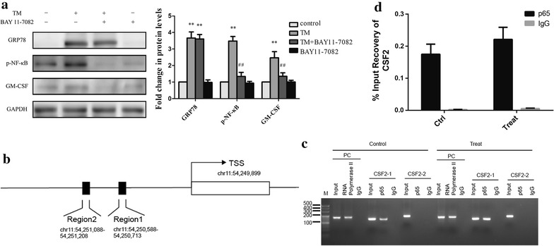
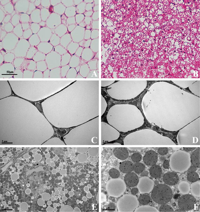
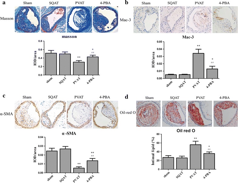
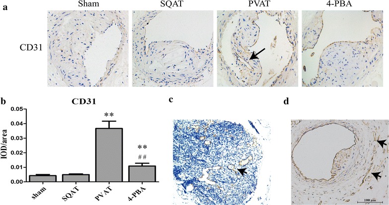
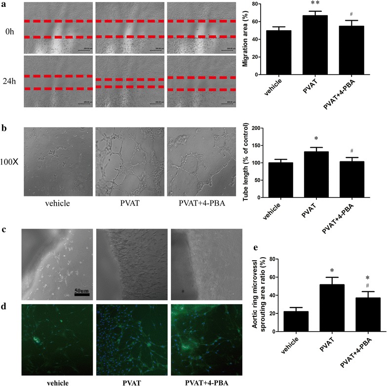
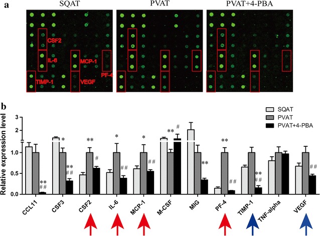
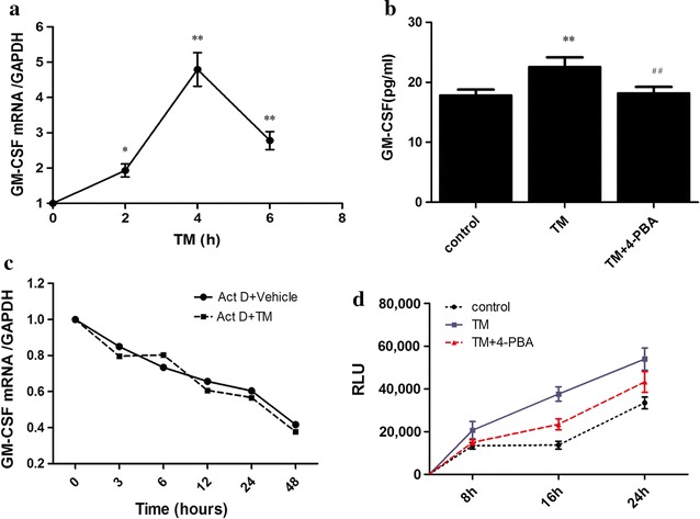
Ultrastructural detection by transmission electron microscopy showed transplanted PVAT was a mixed population of white and brown adipocytes with abundant mitochondria. Transplanted PVAT increased the intraplaque macrophage infiltration, lipid core, intimal and vasa vasorum neovascularization and MMP2/9 expression in plaque while decreased smooth muscle cells and collagen in atherosclerotic plaque, which were restored by local 4-PBA-treatment. Antibody array analysis showed that 4-PBA reduced several angiogenic factors [Granulocyte Macrophage Colony Stimulating Factor (GM-CSF), MCP-1, IL-6] secreted by PVAT. Besides, conditioned medium from 4-PBA treated-PVAT inhibited tube formation and migration capacity of endothelial cells and ex vivo mouse aortic ring angiogenesis compared to conditioned medium from transplanted PVAT. mRNA expression and protein levels of GM-CSF were markedly elevated in adipocytes under ER stress which would be suppressed by 4-PBA. In addition, ER stress enhanced NF-κB binding to the promoter of the mouse GM-CSF gene in adipocytes confirmed by Chromatin immunoprecipitation analyses.
Our findings demonstrate that ER stress in PVAT destabilizes atherosclerotic plaque, in part through increasing GM-CSF paracrine via transcription factor NF-κB.
血管周围脂肪组织(PVAT)加速斑块进展并增加心血管风险。我们检验了这样一个假说,即 PVAT 有助于斑块易损性,并研究了 PVAT 中的内质网应激(ER 应激)是否在易损斑块中发挥重要作用。
我们从供体小鼠中取出颈总动脉套管 6 周后,将胸主动脉 PVAT 或皮下脂肪组织(作为对照)移植到受体载脂蛋白 E 缺陷(apoE)小鼠的颈动脉中。移植后 2 周,局部给予 ER 应激抑制剂 4-苯丁酸(4-PBA)至移植的 PVAT,4 周后处死动物。通过免疫组织化学定量斑块成分和新生血管形成。使用小鼠血管生成抗体阵列试剂盒检测移植脂肪组织产生的血管生成因子。体外管形成试验、划痕伤口迁移试验和小鼠主动脉环试验用于评估移植的 PVAT 上清液的血管生成能力。
透射电子显微镜的超微结构检测显示,移植的 PVAT 是一种含有丰富线粒体的白色和棕色脂肪细胞的混合群体。移植的 PVAT 增加了斑块内的巨噬细胞浸润、脂质核心、内膜和血管腔的新生血管形成以及 MMP2/9 的表达,同时减少了动脉粥样硬化斑块中的平滑肌细胞和胶原,而这些变化可通过局部 4-PBA 治疗得到恢复。抗体阵列分析表明,4-PBA 降低了 PVAT 分泌的几种血管生成因子[粒细胞巨噬细胞集落刺激因子(GM-CSF)、单核细胞趋化蛋白-1(MCP-1)、白细胞介素-6]。此外,与移植的 PVAT 相比,经 4-PBA 处理的 PVAT 的条件培养基抑制了内皮细胞的管形成和迁移能力以及离体小鼠主动脉环的血管生成。在 ER 应激下,脂肪细胞中的 GM-CSF mRNA 表达和蛋白水平显著升高,而 4-PBA 可抑制其表达。此外,染色质免疫沉淀分析证实,ER 应激增强了 NF-κB 与脂肪细胞中 GM-CSF 基因启动子的结合。
我们的研究结果表明,PVAT 中的 ER 应激通过转录因子 NF-κB 增加 GM-CSF 的旁分泌作用来破坏动脉粥样硬化斑块的稳定性。