Department of Molecular Life Sciences, University of Zurich, Winterthurerstrasse 190, CH-8057 Zurich, Switzerland.
MRC Laboratory for Molecular Cell Biology, University College London, Gower St., London WC1E 6BT, UK.
Viruses. 2018 Apr 18;10(4):202. doi: 10.3390/v10040202.
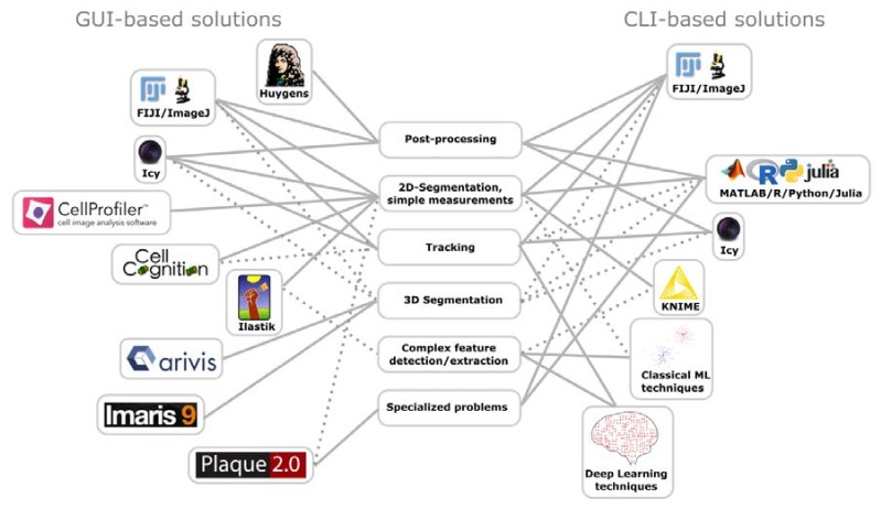
Viruses threaten humans, livestock, and plants, and are difficult to combat. Imaging of viruses by light microscopy is key to uncover the nature of known and emerging viruses in the quest for finding new ways to treat viral disease and deepening the understanding of virus–host interactions. Here, we provide an overview of recent technology for imaging cells and viruses by light microscopy, in particular fluorescence microscopy in static and live-cell modes. The review lays out guidelines for how novel fluorescent chemical probes and proteins can be used in light microscopy to illuminate cells, and how they can be used to study virus infections. We discuss advantages and opportunities of confocal and multi-photon microscopy, selective plane illumination microscopy, and super-resolution microscopy. We emphasize the prevalent concepts in image processing and data analyses, and provide an outlook into label-free digital holographic microscopy for virus research.
病毒威胁着人类、牲畜和植物,并且难以防治。通过光学显微镜对病毒进行成像,是揭示已知和新兴病毒本质的关键,也是寻找治疗病毒性疾病的新方法和加深对病毒-宿主相互作用理解的关键。在这里,我们提供了一个关于通过光学显微镜对细胞和病毒进行成像的最新技术概述,特别是在静态和活细胞模式下的荧光显微镜。该综述阐述了如何在光学显微镜中使用新型荧光化学探针和蛋白质来照亮细胞,以及如何使用它们来研究病毒感染。我们讨论了共聚焦和多光子显微镜、选择性平面照明显微镜和超分辨率显微镜的优势和机会。我们强调了图像处理和数据分析中的流行概念,并对用于病毒研究的无标记数字全息显微镜进行了展望。