Zlabinger Katrin, Lukovic Dominika, Hemetsberger Rayyan, Gugerell Alfred, Winkler Johannes, Mandic Ljubica, Traxler Denise, Spannbauer Andreas, Wolbank Susanne, Zanoni Gerald, Kaun Christoph, Posa Aniko, Gyenes Andrea, Petrasi Zsolt, Petnehazy Örs, Repa Imre, Hofer-Warbinek Renate, de Martin Rainer, Gruber Florian, Charwat Silvia, Huber Kurt, Pavo Noemi, Pavo Imre J, Nyolczas Noemi, Kraitchman Dara L, Gyöngyösi Mariann
Department of Cardiology, Medical University of Vienna, Vienna, Austria.
Ludwig Boltzmann Institute for Clinical and Experimental Traumatology/AUVA Research Center Austrian Cluster for Tissue Regeneration, Vienna, Austria.
Front Bioeng Biotechnol. 2018 Apr 4;6:35. doi: 10.3389/fbioe.2018.00035. eCollection 2018.
Intracoronary (IC) injection of mesenchymal stem cells (MSCs) results in a prompt decrease of absolute myocardial blood flow (AMF) with late and incomplete recovery of myocardial tissue perfusion. Here, we investigated the effect of decreased AMF on oxidative stress marker matrix metalloproteinase-2 (MMP-2) and its influence on the fate and homing and paracrine character of MSCs after IC or intramyocardial cell delivery in a closed-chest reperfused myocardial infarction (MI) model in pigs.
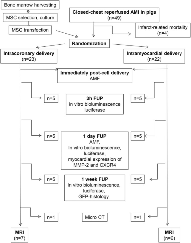
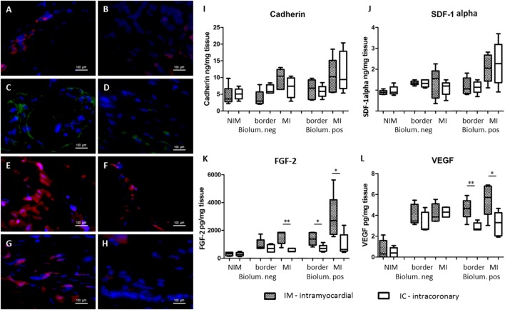
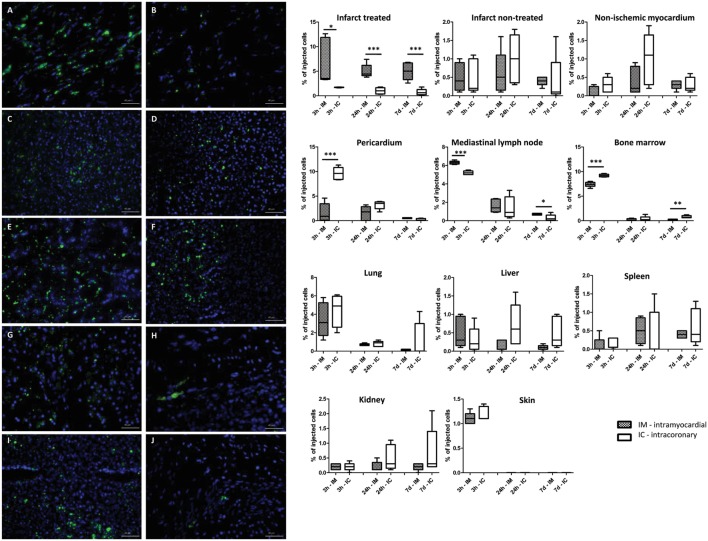
Porcine MSCs were transiently transfected with Ad-Luc and Ad-green fluorescent protein (GFP). One week after MI, the GFP-Luc-MSCs were injected either IC (group IC, 11.00 ± 1.07 × 10) or intramyocardially (group IM, 9.88 ± 1.44 × 10). AMF was measured before, immediately after, and 24 h post GFP-Luc-MSC delivery. bioluminescence signal was used to identify tissue samples containing GFP-Luc-MSCs. Myocardial tissue MMP-2 and CXCR4 receptor expression (index of homing signal) were measured in bioluminescence positive and negative infarcted and border, and non-ischemic myocardial areas 1-day post cell transfer. At 7-day follow-up, myocardial homing (cadherin, CXCR4, and stromal derived factor-1alpha) and angiogenic [fibroblast growth factor 2 (FGF2) and VEGF] were quantified by ELISA of homogenized myocardial tissues from the bioluminescence positive and negative infarcted and border, and non-ischemic myocardium. Biodistribution of the implanted cells was quantified by using Luciferase assay and confirmed by fluorescence immunochemistry. Global left ventricular ejection fraction (LVEF) was measured at baseline and 1-month post cell therapy using magnet resonance image.
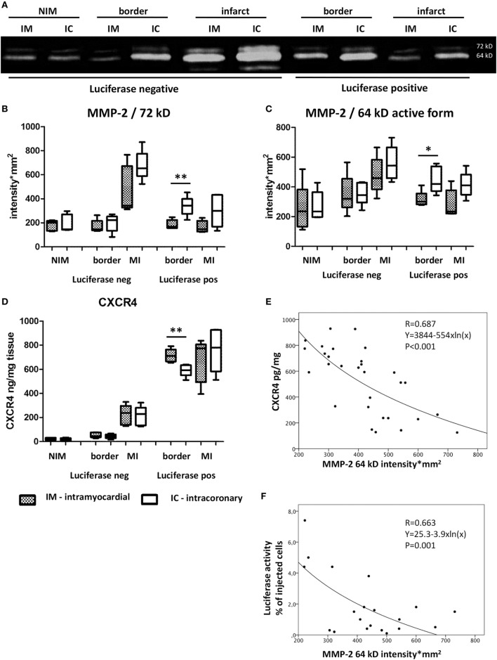
AMF decreased immediately after IC cell delivery, while no change in tissue perfusion was found in the IM group (42.6 ± 11.7 vs. 56.9 ± 16.7 ml/min, = 0.018). IC delivery led to a significant increase in myocardial MMP-2 64 kD expression (448 ± 88 vs. 315 ± 54 intensity × mm, = 0.021), and decreased expression of CXCR4 (592 ± 50 vs. 714 ± 54 pg/tissue/ml, = 0.006), with significant exponential decay between MMP-2 and CXCR4 ( = 0.679, < 0.001). FGF2 and VEGF of the bioluminescence infarcted and border zone of homogenized tissues were significantly elevated in the IM goups as compared to IC group. LVEF increase was significantly higher in IM group (0.8 ± 8.4 vs 5.3 ± 5.2%, = 0.046) at the 1-month follow up.
Intracoronary stem cell delivery decreased AMF, with consequent increase in myocardial expression of MMP-2 and reduced CXCR4 expression with lower level of myocardial homing and angiogenic factor release as compared to IM cell delivery.
冠状动脉内(IC)注射间充质干细胞(MSC)可导致心肌绝对血流量(AMF)迅速下降,且心肌组织灌注恢复延迟且不完全。在此,我们在猪的闭胸再灌注心肌梗死(MI)模型中,研究了AMF降低对氧化应激标志物基质金属蛋白酶-2(MMP-2)的影响,以及IC或心肌内细胞递送后其对MSC的命运、归巢和旁分泌特性的影响。
用腺病毒载体介导的荧光素酶(Ad-Luc)和绿色荧光蛋白(Ad-GFP)对猪MSC进行瞬时转染。MI后1周,将绿色荧光蛋白-荧光素酶-MSC分别经IC(IC组,11.00±1.07×10)或心肌内(IM组,9.88±1.44×10)注射。在注射绿色荧光蛋白-荧光素酶-MSC前、注射后即刻及注射后24小时测量AMF。利用生物发光信号识别含有绿色荧光蛋白-荧光素酶-MSC的组织样本。在细胞转移后1天,测量生物发光阳性和阴性梗死区、边缘区及非缺血心肌区域的心肌组织MMP-2和CXCR4受体表达(归巢信号指数)。在7天随访时,通过酶联免疫吸附测定法(ELISA)对生物发光阳性和阴性梗死区、边缘区及非缺血心肌的匀浆心肌组织中的心肌归巢相关因子(钙黏蛋白、CXCR4和基质细胞衍生因子-1α)和血管生成相关因子[成纤维细胞生长因子2(FGF2)和血管内皮生长因子(VEGF)]进行定量分析。通过荧光素酶测定法定量植入细胞的生物分布,并通过荧光免疫化学法进行确认。使用磁共振成像在基线及细胞治疗后1个月测量整体左心室射血分数(LVEF)。
IC组细胞注射后AMF即刻下降,而IM组组织灌注无变化(42.6±11.7 vs. 56.9±16.7 ml/min,P = 0.018)。IC组注射导致心肌MMP-2 64 kD表达显著增加(448±88 vs. 315±54光密度×mm,P = 0.021),CXCR4表达降低(592±50 vs. 714±54 pg/组织/ml,P = 0.006),MMP-2与CXCR4之间呈显著指数衰减关系(r = 0.679,P < 0.001)。与IC组相比,IM组匀浆组织生物发光梗死区和边缘区的FGF2和VEGF显著升高。在1个月随访时,IM组LVEF增加显著高于IC组(0.8±8.4 vs 5.3±5.2%,P = 0.046)。
与心肌内细胞递送相比,冠状动脉内干细胞递送降低了AMF,导致心肌MMP-2表达增加、CXCR4表达降低,心肌归巢和血管生成因子释放水平降低。