Tumor Microenvironment Laboratory, QIMR Berghofer Medical Research Institute, Herston, Australia.
Werner Siemens Imaging Center, Department of Preclinical Imaging and Radiopharmacy, Eberhard Karls University Tübingen, Tübingen, Germany.
PLoS One. 2018 Apr 20;13(4):e0196040. doi: 10.1371/journal.pone.0196040. eCollection 2018.
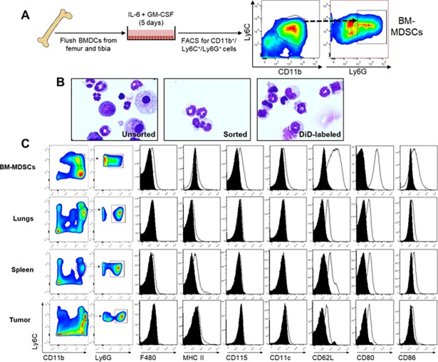
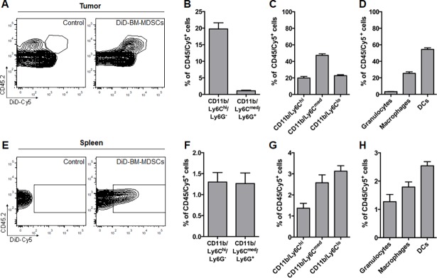
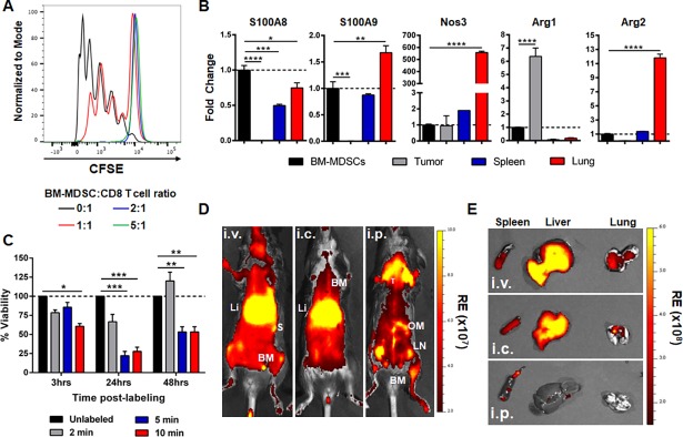
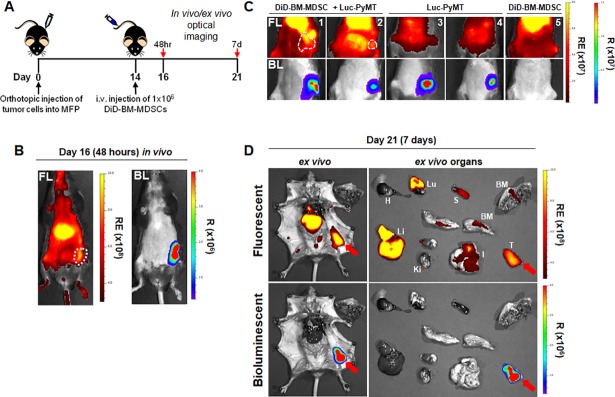
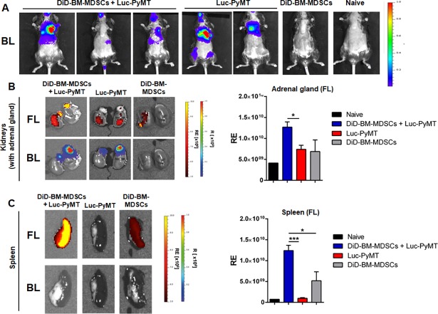
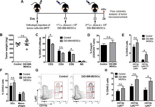
Myeloid-derived suppressor cells (MDSCs) are a heterogeneous population of immature myeloid progenitor cells that are expanded in cancer and act as potent suppressors of the anti-tumor immune response. MDSCs consist of two major subsets, namely monocytic (M-) MDSCs and granulocytic (G-) MDSCs that differ with respect to their phenotype, morphology and mechanisms of suppression. Here, we cultured bone marrow cells with IL-6 and GM-CSF in vitro to generate a population of bone marrow MDSCs (BM-MDSCs) similar to G-MDSCs from tumor-bearing mice in regards to phenotype, morphology and suppressive-function. Through fluorescent labeling of these BM-MDSCs and optical imaging, we could visualize the recruitment and localization of BM-MDSCs in breast tumor-bearing mice in vivo. Furthermore, we were able to demonstrate that BM-MDSCs home to primary and metastatic breast tumors, but have no significant effect on tumor growth or progression. Ex vivo flow cytometry characterization of BM-MDSCs after adoptive transfer demonstrated both organ-and tumor-specific effects on their phenotype and differentiation, demonstrating the importance of the local microenvironment on MDSC fate and function. In this study, we have developed a method to generate, visualize and detect BM-MDSCs in vivo and ex vivo through optical imaging and flow cytometry, in order to understand the organ-specific changes rendered to MDSCs in breast cancer.
髓系来源的抑制细胞(MDSCs)是一种异质性的未成熟髓系祖细胞群体,在癌症中扩增,并作为抗肿瘤免疫反应的有效抑制物。MDSCs 由两个主要亚群组成,即单核细胞(M-)MDSCs 和粒细胞(G-)MDSCs,它们在表型、形态和抑制机制方面存在差异。在这里,我们通过体外培养骨髓细胞与 IL-6 和 GM-CSF,生成了一群类似于荷瘤小鼠来源的 G-MDSCs 的骨髓 MDSCs(BM-MDSCs),在表型、形态和抑制功能方面都相似。通过对这些 BM-MDSCs 进行荧光标记和光学成像,我们可以在体内可视化 BM-MDSCs 在乳腺癌荷瘤小鼠中的募集和定位。此外,我们还能够证明 BM-MDSCs 归巢到原发性和转移性乳腺癌肿瘤,但对肿瘤生长或进展没有显著影响。过继转移后对 BM-MDSCs 的体外流式细胞术特征分析表明,它们的表型和分化具有器官和肿瘤特异性效应,这表明局部微环境对 MDSC 命运和功能的重要性。在这项研究中,我们开发了一种通过光学成像和流式细胞术在体内和体外生成、可视化和检测 BM-MDSCs 的方法,以了解乳腺癌中 MDSCs 所呈现的器官特异性变化。