Goller-Bulut D, Özcan G, Avci F
Department of Oral and Maxillofacial Radiology, Faculty of Dentistry Abant İzzet Baysal University, 14000, Bolu, Turkey,
Med Oral Patol Oral Cir Bucal. 2018 May 1;23(3):e282-e289. doi: 10.4317/medoral.22274.
The aim of this retrospective study was to compare the morphological features of neurovascular canals and foramina of patients with medication-related osteonecrosis of the jaws (MRONJ) and healthy individuals by using cone beam computed tomography (CBCT).
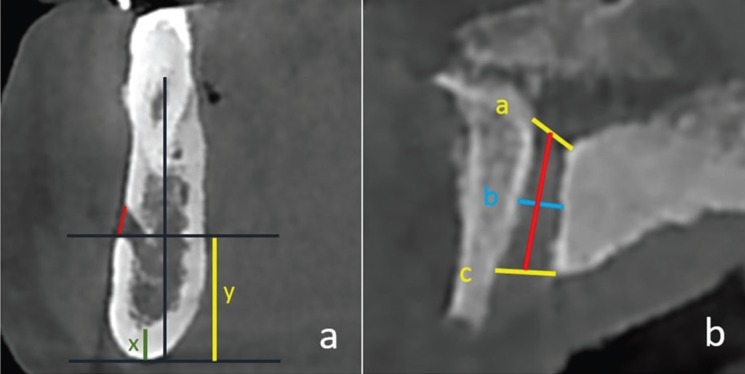
The CBCT images of 58 patients under bisphosphonate therapy diagnosed with MRONJ and age gender- matched controls were retrospectively evaluated. The diameter of mandibular and nasopalatine canal and mandibular, mental and lingual foramina were measured on several sections of CBCT. The value of mental index (MI) and panoramic mandibular index (PMI) were also assessed.
The mean value of diametric measurements for all neurovascular canals and foramina in MRONJ patients were narrower than controls. Left mandibular foramen was the most affected area (p<0.001). There were significantly difference in all measurements of mental foramen, lingual foramen and mandibular incisive canal between two groups (p<0.05). PMI of MRONJ subjects were also significantly differences in both sides (p<0.05).
In MRONJ patient, neurovascular canals and foramina are affected due to the alterations in bone remodeling. Therefore, the diametric measurement of neurovascular canals and assessment of MI and PMI on CBCT, is a potentially useful method for detection of early changes associated with bisphosphonate therapy and for predict areas where new necrosis may occur.
本回顾性研究的目的是通过使用锥形束计算机断层扫描(CBCT)比较药物性颌骨坏死(MRONJ)患者和健康个体的神经血管管及孔的形态特征。
回顾性评估58例诊断为MRONJ且正在接受双膦酸盐治疗的患者以及年龄和性别匹配的对照组的CBCT图像。在CBCT的多个层面上测量下颌管、鼻腭管以及下颌孔、颏孔和舌孔的直径。还评估了颏指数(MI)和全景下颌指数(PMI)的值。
MRONJ患者所有神经血管管及孔的直径测量平均值均比对照组窄。左侧下颌孔是受影响最严重的区域(p<0.001)。两组之间颏孔、舌孔和下颌切牙管的所有测量值均存在显著差异(p<0.05)。MRONJ受试者的PMI两侧也存在显著差异(p<0.05)。
在MRONJ患者中,神经血管管及孔因骨重塑改变而受到影响。因此,在CBCT上对神经血管管进行直径测量以及评估MI和PMI,是检测与双膦酸盐治疗相关的早期变化以及预测可能发生新坏死区域的一种潜在有用方法。