• 文献检索
  • 文档翻译
  • 深度研究
  • 学术资讯
  • Suppr Zotero 插件Zotero 插件
  • 邀请有礼
  • 套餐&价格
  • 历史记录
应用&插件
Suppr Zotero 插件Zotero 插件浏览器插件Mac 客户端Windows 客户端微信小程序
定价
高级版会员购买积分包购买API积分包
服务
文献检索文档翻译深度研究API 文档MCP 服务
关于我们
关于 Suppr公司介绍联系我们用户协议隐私条款
关注我们

Suppr 超能文献

核心技术专利:CN118964589B侵权必究
粤ICP备2023148730 号-1Suppr @ 2026

文献检索

告别复杂PubMed语法,用中文像聊天一样搜索,搜遍4000万医学文献。AI智能推荐,让科研检索更轻松。

立即免费搜索

文件翻译

保留排版,准确专业,支持PDF/Word/PPT等文件格式,支持 12+语言互译。

免费翻译文档

深度研究

AI帮你快速写综述,25分钟生成高质量综述,智能提取关键信息,辅助科研写作。

立即免费体验

三维组织学中组织重建算法的比较分析。

Comparative analysis of tissue reconstruction algorithms for 3D histology.

机构信息

Faculty of Medicine and Life Sciences, University of Tampere, Tampere, Finland.

Faculty of Biomedical Sciences and Engineering, Tampere University of Technology, Tampere, Finland.

出版信息

Bioinformatics. 2018 Sep 1;34(17):3013-3021. doi: 10.1093/bioinformatics/bty210.

DOI:10.1093/bioinformatics/bty210
PMID:29684099
原文链接:https://pmc.ncbi.nlm.nih.gov/articles/PMC6129300/
Abstract

MOTIVATION

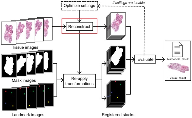
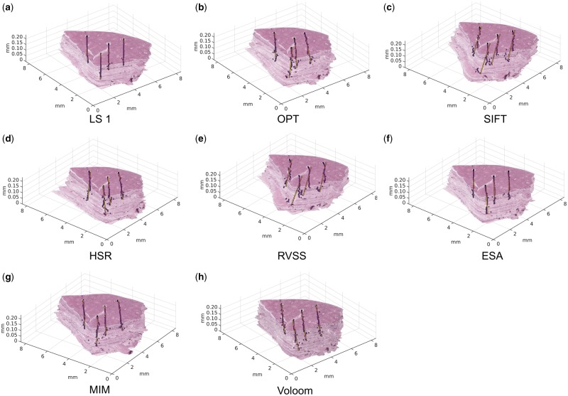
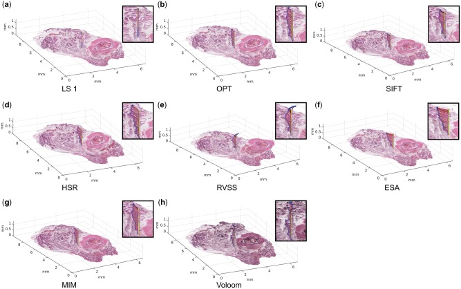
Digital pathology enables new approaches that expand beyond storage, visualization or analysis of histological samples in digital format. One novel opportunity is 3D histology, where a three-dimensional reconstruction of the sample is formed computationally based on serial tissue sections. This allows examining tissue architecture in 3D, for example, for diagnostic purposes. Importantly, 3D histology enables joint mapping of cellular morphology with spatially resolved omics data in the true 3D context of the tissue at microscopic resolution. Several algorithms have been proposed for the reconstruction task, but a quantitative comparison of their accuracy is lacking.

RESULTS

We developed a benchmarking framework to evaluate the accuracy of several free and commercial 3D reconstruction methods using two whole slide image datasets. The results provide a solid basis for further development and application of 3D histology algorithms and indicate that methods capable of compensating for local tissue deformation are superior to simpler approaches.

AVAILABILITY AND IMPLEMENTATION

Code: https://github.com/BioimageInformaticsTampere/RegBenchmark. Whole slide image datasets: http://urn.fi/urn: nbn: fi: csc-kata20170705131652639702.

SUPPLEMENTARY INFORMATION

Supplementary data are available at Bioinformatics online.

摘要

动机

数字病理学能够实现新的方法,超越数字格式的组织样本存储、可视化或分析。一个新颖的机会是三维组织学,其中根据组织切片的序列进行计算形成样本的三维重建。这允许在三维空间中检查组织架构,例如用于诊断目的。重要的是,三维组织学能够在组织的真实三维环境中,以微观分辨率联合映射细胞形态与空间分辨的组学数据。已经提出了几种用于重建任务的算法,但缺乏对其准确性的定量比较。

结果

我们开发了一个基准测试框架,使用两个全幻灯片图像数据集来评估几种免费和商业 3D 重建方法的准确性。结果为进一步开发和应用三维组织学算法提供了坚实的基础,并表明能够补偿局部组织变形的方法优于更简单的方法。

可用性和实现

代码:https://github.com/BioimageInformaticsTampere/RegBenchmark。全幻灯片图像数据集:http://urn.fi/urn: nbn: fi: csc-kata20170705131652639702。

补充信息

补充数据可在 Bioinformatics 在线获得。

https://cdn.ncbi.nlm.nih.gov/pmc/blobs/f598/6129300/7bcb01cd7efe/bty210f3.jpg
https://cdn.ncbi.nlm.nih.gov/pmc/blobs/f598/6129300/343236891d98/bty210f1.jpg
https://cdn.ncbi.nlm.nih.gov/pmc/blobs/f598/6129300/07bd6b3fef37/bty210f2.jpg
https://cdn.ncbi.nlm.nih.gov/pmc/blobs/f598/6129300/7bcb01cd7efe/bty210f3.jpg
https://cdn.ncbi.nlm.nih.gov/pmc/blobs/f598/6129300/343236891d98/bty210f1.jpg
https://cdn.ncbi.nlm.nih.gov/pmc/blobs/f598/6129300/07bd6b3fef37/bty210f2.jpg
https://cdn.ncbi.nlm.nih.gov/pmc/blobs/f598/6129300/7bcb01cd7efe/bty210f3.jpg

相似文献

1
Comparative analysis of tissue reconstruction algorithms for 3D histology.三维组织学中组织重建算法的比较分析。
Bioinformatics. 2018 Sep 1;34(17):3013-3021. doi: 10.1093/bioinformatics/bty210.
2
3D volume reconstruction from serial breast specimen radiographs for mapping between histology and 3D whole specimen imaging.通过乳腺标本连续X光片进行3D体积重建,用于组织学与3D全标本成像之间的映射。
Med Phys. 2017 Mar;44(3):935-948. doi: 10.1002/mp.12077.
3
Framework for 3D histologic reconstruction and fusion with in vivo MRI: Preliminary results of characterizing pulmonary inflammation in a mouse model.用于三维组织学重建及与活体磁共振成像融合的框架:小鼠模型中肺部炎症特征化的初步结果
Med Phys. 2015 Aug;42(8):4822-32. doi: 10.1118/1.4923161.
4
A Survey of Methods for 3D Histology Reconstruction.三维组织学重建方法研究综述。
Med Image Anal. 2018 May;46:73-105. doi: 10.1016/j.media.2018.02.004. Epub 2018 Feb 21.
5
Three-dimensional reconstruction with serial whole-mount sections of oral tongue squamous cell carcinoma: A preliminary study.舌鳞状细胞癌连续全器官切片的三维重建:初步研究。
J Oral Pathol Med. 2018 Jan;47(1):53-59. doi: 10.1111/jop.12644. Epub 2017 Oct 23.
6
Image registration of ex-vivo MRI to sparsely sectioned histology of hippocampal and neocortical temporal lobe specimens.将离体 MRI 与海马和新皮质颞叶标本的稀疏切片组织学图像进行配准。
Neuroimage. 2013 Dec;83:770-81. doi: 10.1016/j.neuroimage.2013.07.053. Epub 2013 Jul 26.
7
Showing their true colors: a practical approach to volume rendering from serial sections.展现真面目:一种从连续切片进行体绘制的实用方法。
BMC Dev Biol. 2010 Apr 21;10:41. doi: 10.1186/1471-213X-10-41.
8
Fast 3D iterative image reconstruction for SPECT with rotating slat collimators.用于配备旋转板准直器的单光子发射计算机断层扫描(SPECT)的快速三维迭代图像重建
Phys Med Biol. 2009 Feb 7;54(3):715-29. doi: 10.1088/0031-9155/54/3/016. Epub 2009 Jan 9.
9
3-dimensional digital reconstruction of the murine coronary system for the evaluation of chronic allograft vasculopathy.用于评估慢性移植物血管病的小鼠冠状动脉系统的三维数字重建
Diagn Pathol. 2015 Mar 28;10:16. doi: 10.1186/s13000-015-0248-6.
10
3D prostate histology reconstruction: an evaluation of image-based and fiducial-based algorithms.3D 前列腺组织学重建:基于图像和基于标记的算法评估。
Med Phys. 2013 Sep;40(9):093501. doi: 10.1118/1.4816946.

引用本文的文献

1
3D visualization of uterus and ovary: tissue clearing techniques and biomedical applications.子宫和卵巢的三维可视化:组织透明化技术及生物医学应用
Front Bioeng Biotechnol. 2025 Jul 7;13:1610539. doi: 10.3389/fbioe.2025.1610539. eCollection 2025.
2
Spatial landmark detection and tissue registration with deep learning.基于深度学习的空间地标检测和组织配准。
Nat Methods. 2024 Apr;21(4):673-679. doi: 10.1038/s41592-024-02199-5. Epub 2024 Mar 4.
3
Three-dimensional histology reveals dissociable human hippocampal long-axis gradients of Alzheimer's pathology.

本文引用的文献

1
Transformation diffusion reconstruction of three-dimensional histology volumes from two-dimensional image stacks.从二维图像堆叠中重建三维组织学体积的变换扩散。
Med Image Anal. 2017 May;38:184-204. doi: 10.1016/j.media.2017.03.004. Epub 2017 Mar 23.
2
Virtual reality assisted microscopy data visualization and colocalization analysis.虚拟现实辅助显微镜数据可视化与共定位分析。
BMC Bioinformatics. 2017 Feb 15;18(Suppl 2):64. doi: 10.1186/s12859-016-1446-2.
3
The race to map the human body - one cell at a time.一次一个细胞地绘制人体图谱的竞赛。
三维组织学揭示了可分离的人类海马体阿尔茨海默病病理的长轴梯度。
Alzheimers Dement. 2024 Apr;20(4):2606-2619. doi: 10.1002/alz.13695. Epub 2024 Feb 18.
4
3D visualization and printing: An "Anatomical Engineering" trend revealing underlying morphology via innovation and reconstruction towards future of veterinary anatomy.三维可视化和打印:一种“解剖工程”趋势,通过创新和重建揭示潜在形态,展望未来的兽医解剖学。
Anat Sci Int. 2024 Mar;99(2):159-182. doi: 10.1007/s12565-023-00755-1. Epub 2024 Jan 18.
5
Tissue clearing and imaging approaches for in toto analysis of the reproductive system†.组织透明化和成像方法用于生殖系统的整体分析。
Biol Reprod. 2024 Jun 12;110(6):1041-1054. doi: 10.1093/biolre/ioad182.
6
Comparison of consecutive and restained sections for image registration in histopathology.用于组织病理学图像配准的连续切片与重新染色切片的比较。
J Med Imaging (Bellingham). 2023 Nov;10(6):067501. doi: 10.1117/1.JMI.10.6.067501. Epub 2023 Nov 30.
7
Decoding the tumor microenvironment with spatial technologies.利用空间技术解码肿瘤微环境。
Nat Immunol. 2023 Dec;24(12):1982-1993. doi: 10.1038/s41590-023-01678-9. Epub 2023 Nov 27.
8
A Multi-Stain Breast Cancer Histological Whole-Slide-Image Data Set from Routine Diagnostics.多染色乳腺癌组织学全切片图像数据集来自常规诊断。
Sci Data. 2023 Aug 24;10(1):562. doi: 10.1038/s41597-023-02422-6.
9
Virtual alignment of pathology image series for multi-gigapixel whole slide images.多吉帕像素全玻片图像的病理学图像系列的虚拟配准。
Nat Commun. 2023 Jul 26;14(1):4502. doi: 10.1038/s41467-023-40218-9.
10
Deep learning-based diagnosis of feline hypertrophic cardiomyopathy.基于深度学习的猫肥厚型心肌病诊断。
PLoS One. 2023 Feb 2;18(2):e0280438. doi: 10.1371/journal.pone.0280438. eCollection 2023.
Nature. 2017 Feb 20;542(7642):404-405. doi: 10.1038/nature.2017.21508.
4
Algorithm sensitivity analysis and parameter tuning for tissue image segmentation pipelines.用于组织图像分割流程的算法敏感性分析和参数调整
Bioinformatics. 2017 Apr 1;33(7):1064-1072. doi: 10.1093/bioinformatics/btw749.
5
Digital pathology in clinical use: where are we now and what is holding us back?临床应用中的数字病理学:我们目前的状况以及阻碍我们发展的因素是什么?
Histopathology. 2017 Jan;70(1):134-145. doi: 10.1111/his.12993.
6
Imagining the future of bioimage analysis.畅想生物图像分析的未来。
Nat Biotechnol. 2016 Dec 7;34(12):1250-1255. doi: 10.1038/nbt.3722.
7
ImageJ-MATLAB: a bidirectional framework for scientific image analysis interoperability.ImageJ-MATLAB:用于科学图像分析互操作性的双向框架。
Bioinformatics. 2017 Feb 15;33(4):629-630. doi: 10.1093/bioinformatics/btw681.
8
Visualization and analysis of gene expression in tissue sections by spatial transcriptomics.空间转录组学在组织切片中基因表达的可视化和分析。
Science. 2016 Jul 1;353(6294):78-82. doi: 10.1126/science.aaf2403.
9
Methodology to study the three-dimensional spatial distribution of prostate cancer and their dependence on clinical parameters.研究前列腺癌三维空间分布及其对临床参数依赖性的方法。
J Med Imaging (Bellingham). 2015 Jul;2(3):037502. doi: 10.1117/1.JMI.2.3.037502. Epub 2015 Jul 29.
10
Three-dimensional immersive virtual reality for studying cellular compartments in 3D models from EM preparations of neural tissues.用于研究神经组织电子显微镜制备的三维模型中细胞区室的三维沉浸式虚拟现实技术。
J Comp Neurol. 2016 Jan 1;524(1):23-38. doi: 10.1002/cne.23852. Epub 2015 Aug 11.