Department of Anesthesiology and the Center for Shock, Trauma and Anesthesiology Research (STAR), University of Maryland School of Medicine, Baltimore, MD, USA.
Department of Orthopaedics, University of Maryland School of Medicine, Baltimore, MD, USA.
Cell Death Dis. 2018 May 1;9(5):476. doi: 10.1038/s41419-018-0469-1.
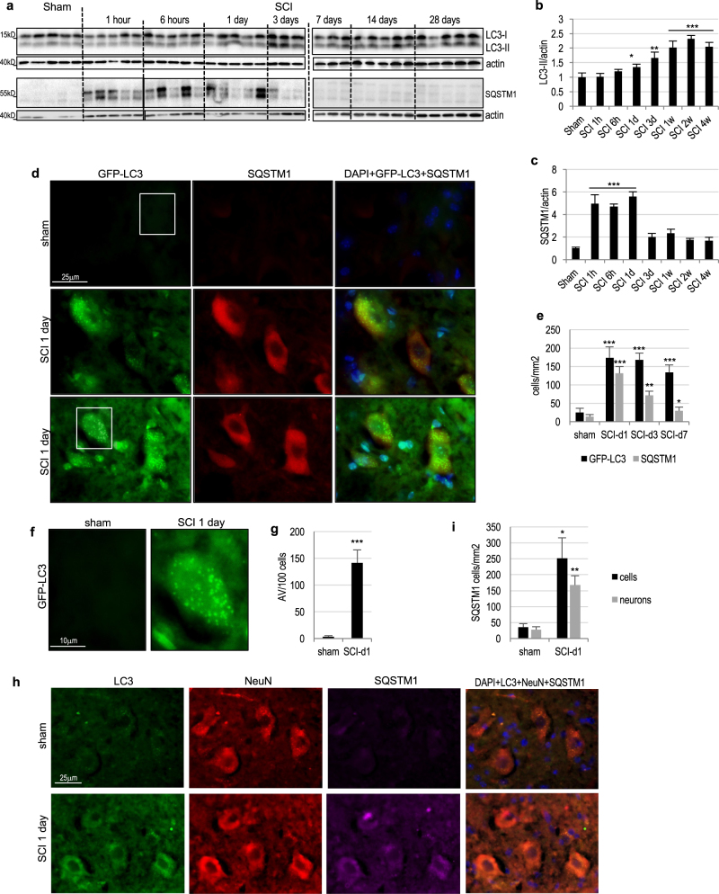
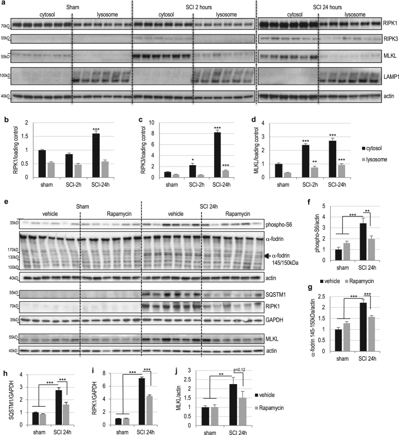
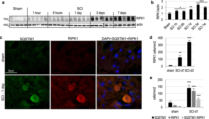
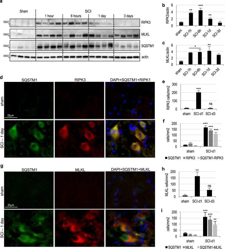
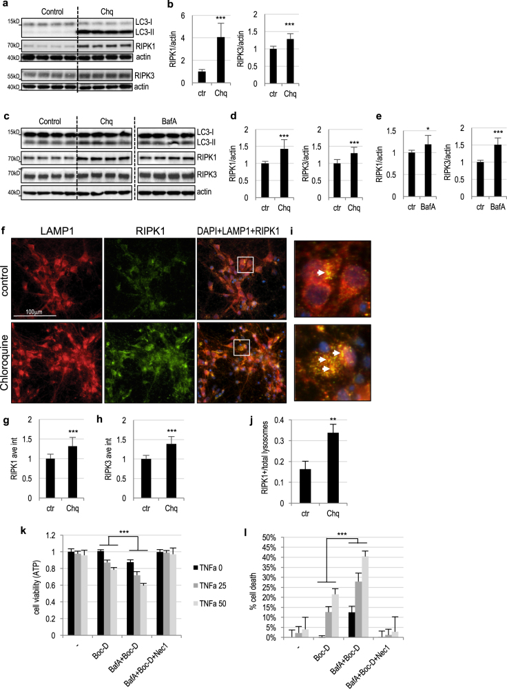
Necroptosis, a regulated necrosis pathway mediated by the receptor-interacting protein kinases 1 and 3 (RIPK1 and RIPK3), is induced following spinal cord injury (SCI) and thought to contribute to neuronal and glial cell death. However, mechanisms leading to activation of necroptosis after SCI remain unclear. We have previously shown that autophagy, a catabolic pathway facilitating degradation of cytoplasmic proteins and organelles in a lysosome-dependent manner, is inhibited following SCI in rats. Our current data confirm that inhibition of autophagy also occurs after thoracic contusive SCI in the mouse model, as indicated by accumulation of both the autophagosome marker, LC3-II and autophagy cargo protein, p62/SQSTM1. This was most pronounced in the ventral horn neurons and was caused by rapid inhibition of lysosomal function after SCI. Interestingly, RIPK1, RIPK3, and the necroptosis effector protein MLKL also rapidly accumulated after SCI and localized to neurons with disrupted autophagy, suggesting that these events may be related. To determine if lysosomal dysfunction could contribute to induction of necroptosis, we treated PC12 cells and primary rat cortical neurons with lysosomal inhibitors. This led to rapid accumulation of RIPK1 and RIPK3, confirming that they are normally degraded by the lysosomal pathway. In PC12 cells lysosomal inhibition also sensitized cells to necroptosis induced by tumor necrosis factor α (TNFα) and caspase inhibitor. Imaging studies confirmed that RIPK1 partially localized to lysosomes in both untreated and lysosomal inhibitor treated cells. Similarly, we detected presence of RIPK1, RIPK3 and MLKL in both cytosol and at lysosomes after SCI in vivo. Furthermore, stimulation of autophagy and lysosomal function with rapamycin treatment led to decreased accumulation of RIPK1 and attenuated cell death after SCI. These data suggest that lysosomal dysfunction after SCI may contribute to both inhibition of autophagy and sensitize cells to necroptosis by promoting RIPK1 and RIPK3 accumulation.
细胞程序性坏死是一种由受体相互作用蛋白激酶 1 和 3(RIPK1 和 RIPK3)介导的调节性细胞坏死途径,在脊髓损伤(SCI)后被诱导,并被认为有助于神经元和神经胶质细胞死亡。然而,SCI 后导致细胞程序性坏死激活的机制仍不清楚。我们之前已经表明,自噬是一种促进溶酶体依赖性细胞质蛋白和细胞器降解的分解代谢途径,在大鼠 SCI 后被抑制。我们目前的数据证实,在小鼠模型的胸段挫伤性 SCI 后,自噬也被抑制,这表现在自噬体标记物 LC3-II 和自噬货物蛋白 p62/SQSTM1 的积累。在腹角神经元中最为明显,这是由于 SCI 后溶酶体功能的快速抑制所致。有趣的是,RIPK1、RIPK3 和细胞程序性坏死效应蛋白 MLKL 在 SCI 后也迅速积累,并定位于自噬受损的神经元,表明这些事件可能相关。为了确定溶酶体功能障碍是否有助于诱导细胞程序性坏死,我们用溶酶体抑制剂处理 PC12 细胞和原代大鼠皮质神经元。这导致 RIPK1 和 RIPK3 的快速积累,证实它们通常通过溶酶体途径降解。在 PC12 细胞中,溶酶体抑制也使细胞对肿瘤坏死因子 α(TNFα)和半胱天冬酶抑制剂诱导的细胞程序性坏死敏感。成像研究证实,RIPK1 在未处理和溶酶体抑制剂处理的细胞中部分定位于溶酶体。同样,我们在体内 SCI 后也检测到 RIPK1、RIPK3 和 MLKL 存在于细胞质和溶酶体中。此外,用雷帕霉素处理刺激自噬和溶酶体功能会导致 RIPK1 的积累减少,并减轻 SCI 后的细胞死亡。这些数据表明,SCI 后溶酶体功能障碍可能通过促进 RIPK1 和 RIPK3 的积累,导致自噬抑制和细胞对细胞程序性坏死敏感。