• 文献检索
  • 文档翻译
  • 深度研究
  • 学术资讯
  • Suppr Zotero 插件Zotero 插件
  • 邀请有礼
  • 套餐&价格
  • 历史记录
应用&插件
Suppr Zotero 插件Zotero 插件浏览器插件Mac 客户端Windows 客户端微信小程序
定价
高级版会员购买积分包购买API积分包
服务
文献检索文档翻译深度研究API 文档MCP 服务
关于我们
关于 Suppr公司介绍联系我们用户协议隐私条款
关注我们

Suppr 超能文献

核心技术专利:CN118964589B侵权必究
粤ICP备2023148730 号-1Suppr @ 2026

文献检索

告别复杂PubMed语法,用中文像聊天一样搜索,搜遍4000万医学文献。AI智能推荐,让科研检索更轻松。

立即免费搜索

文件翻译

保留排版,准确专业,支持PDF/Word/PPT等文件格式,支持 12+语言互译。

免费翻译文档

深度研究

AI帮你快速写综述,25分钟生成高质量综述,智能提取关键信息,辅助科研写作。

立即免费体验

下颌前伸固定方法及其对颞下颌关节的影响的有限元分析研究。

Fixation Methods for Mandibular Advancement and Their Effects on Temporomandibular Joint: A Finite Element Analysis Study.

机构信息

Beykent University Vocational School Dental Services, Oral Health Program, Istanbul, Turkey.

Istanbul University Faculty of Dentistry Department of Oral and Maxillofacial Surgery, Istanbul, Turkey.

出版信息

Biomed Res Int. 2020 Feb 21;2020:2810763. doi: 10.1155/2020/2810763. eCollection 2020.

DOI:10.1155/2020/2810763
PMID:32185199
原文链接:https://pmc.ncbi.nlm.nih.gov/articles/PMC7060428/
Abstract

OBJECTIVES

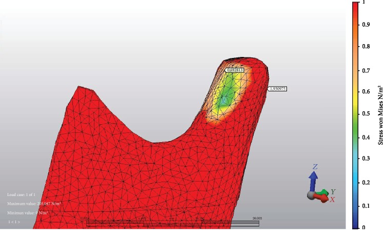
Bilateral sagittal split osteotomy (BSSO) is a common surgical procedure to correct dentofacial deformities that involve the mandible. Usually bicortical bone fixation screw or miniplates with monocortical bone fixation screw were used to gain stability after BSSO. On the other hand, the use of resorbable screw materials had been reported. In this study, our aim is to determine first stress distribution values at the temporomandibular joint (TMJ) and second displacement amounts of each mandibular bone segment.

METHODS

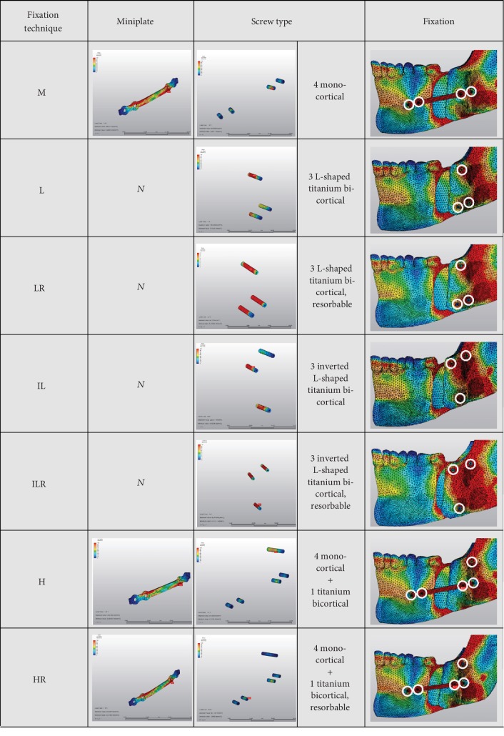
A three-dimensional virtual mesh model of the mandible was constructed. Then, BSSO with 9 mm advancement was simulated using the finite element model (FEM). Fixation between each mandibular segment was also virtually performed using seven different combinations of fixation materials, as follows: miniplate only (M), miniplate and a titanium bicortical bone fixation screw (H), miniplate and a resorbable bicortical bone fixation screw (HR), 3 L-shaped titanium bicortical bone fixation screws (L), 3 L-shaped resorbable bicortical bone fixation screws (LR), 3 inverted L-shaped titanium bicortical bone fixation screws (IL), and 3 inverted L-shaped resorbable bicortical bone fixation screws (ILR).

RESULTS

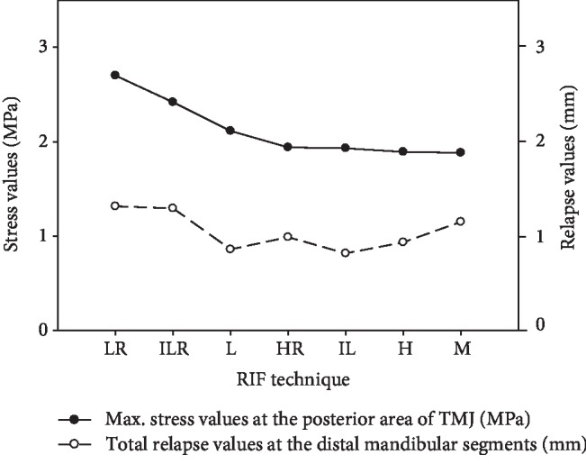
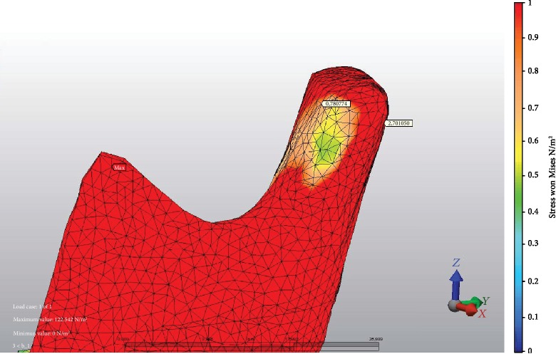
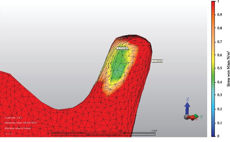
At 9 mm advancement, the biggest stress values at the anterior area TMJ was seen at M fixation and LR fixation at posterior TMJ. The minimum stress values on anterior TMJ were seen at L fixation and M fixation at posterior TMJ. Minimum displacement was seen in IL method. It was followed by L, H, HR, M, ILR, and LR, respectively.

CONCLUSION

According to our results, bicortical screw fixation was associated with more stress on the condyle. In terms of total stress value, especially LR and ILR lead to higher amounts.

摘要

目的

双侧矢状劈开截骨术(BSSO)是一种常见的矫正下颌骨畸形的手术方法。通常使用双皮质骨固定螺钉或带有单皮质骨固定螺钉的微型板在 BSSO 后获得稳定性。另一方面,已经报道了可吸收螺钉材料的使用。在这项研究中,我们的目的是确定第一颞下颌关节(TMJ)的应力分布值和第二下颌骨各段的位移量。

方法

构建了下颌骨的三维虚拟网格模型。然后,使用有限元模型(FEM)模拟了 9mm 的 BSSO 推进。还通过七种不同的固定材料组合虚拟地进行了每个下颌骨段之间的固定,如下所示:仅微型板(M)、微型板和钛双皮质骨固定螺钉(H)、微型板和可吸收双皮质骨固定螺钉(HR)、3 个 L 形钛双皮质骨固定螺钉(L)、3 个 L 形可吸收双皮质骨固定螺钉(LR)、3 个倒置 L 形钛双皮质骨固定螺钉(IL)和 3 个倒置 L 形可吸收双皮质骨固定螺钉(ILR)。

结果

在 9mm 的推进中,在前 TMJ 区域,M 固定和 LR 固定时的最大应力值最大,在后 TMJ 区域,L 固定和 M 固定时的最小应力值最大。在前 TMJ 处的最小位移在 IL 方法中可见。其次是 L、H、HR、M、ILR 和 LR。

结论

根据我们的结果,双皮质螺钉固定与髁突上的更大的应力有关。就总应力值而言,特别是 LR 和 ILR 导致更高的量。

https://cdn.ncbi.nlm.nih.gov/pmc/blobs/47db/7060428/321e435c036d/BMRI2020-2810763.005.jpg
https://cdn.ncbi.nlm.nih.gov/pmc/blobs/47db/7060428/1f3525ed2244/BMRI2020-2810763.001.jpg
https://cdn.ncbi.nlm.nih.gov/pmc/blobs/47db/7060428/72312d61d780/BMRI2020-2810763.002.jpg
https://cdn.ncbi.nlm.nih.gov/pmc/blobs/47db/7060428/59062b4dbc04/BMRI2020-2810763.003.jpg
https://cdn.ncbi.nlm.nih.gov/pmc/blobs/47db/7060428/9658d10f0ce1/BMRI2020-2810763.004.jpg
https://cdn.ncbi.nlm.nih.gov/pmc/blobs/47db/7060428/321e435c036d/BMRI2020-2810763.005.jpg

相似文献

1
Fixation Methods for Mandibular Advancement and Their Effects on Temporomandibular Joint: A Finite Element Analysis Study.下颌前伸固定方法及其对颞下颌关节的影响的有限元分析研究。
Biomed Res Int. 2020 Feb 21;2020:2810763. doi: 10.1155/2020/2810763. eCollection 2020.
2
Does fixation method affects temporomandibular joints after mandibular advancement?固定方式是否会影响下颌前伸术后的颞下颌关节?
J Craniomaxillofac Surg. 2018 Jun;46(6):923-931. doi: 10.1016/j.jcms.2018.03.024. Epub 2018 Apr 7.
3
An in vitro evaluation of rigid internal fixation techniques for sagittal split ramus osteotomies: advancement surgery.下颌升支矢状劈开截骨术的坚固内固定技术的体外评估:前徙手术
J Oral Maxillofac Surg. 2009 Apr;67(4):809-17. doi: 10.1016/j.joms.2008.11.009.
4
Comparison of bicortical, miniplate and hybrid fixation techniques in mandibular advancement and counterclockwise rotation: A finite element analysis study.下颌前徙和逆时针旋转中双皮质、微型钢板及混合固定技术的比较:一项有限元分析研究。
J Stomatol Oral Maxillofac Surg. 2021 Sep;122(4):e7-e14. doi: 10.1016/j.jormas.2021.04.004. Epub 2021 Apr 10.
5
In vitro biomechanical evaluation of fixation methods of sagittal split ramus osteotomy in mandibular setback.下颌后缩矢状劈开下颌支截骨术固定方法的体外生物力学评估
J Craniomaxillofac Surg. 2015 Mar;43(2):186-91. doi: 10.1016/j.jcms.2014.10.023. Epub 2014 Dec 3.
6
Analysis of stress distribution on fixation of bilateral sagittal split ramus osteotomy with resorbable plates and screws using the finite-element method.使用有限元方法分析可吸收接骨板和螺钉固定双侧下颌支矢状劈开截骨术时的应力分布。
J Oral Maxillofac Surg. 2012 Jun;70(6):1434-8. doi: 10.1016/j.joms.2011.05.017. Epub 2011 Aug 6.
7
Comperative evaluation of the current and new design miniplate fixation techniques of the advanced sagittal split ramus osteotomy using three-dimensional finite element analysis.采用三维有限元分析法对当前和新型设计的下颌升支矢状劈开截骨术微型接骨板固定技术的比较评估。
Med Oral Patol Oral Cir Bucal. 2023 Nov 1;28(6):e553-e561. doi: 10.4317/medoral.25964.
8
An in vitro evaluation of rigid internal fixation techniques for sagittal split ramus osteotomies: setback surgery.矢状劈开下颌支截骨术的坚固内固定技术的体外评估:后退手术
J Oral Maxillofac Surg. 2012 Apr;70(4):941-51. doi: 10.1016/j.joms.2011.02.106. Epub 2011 Jul 14.
9
Are bicortical screw and plate osteosynthesis techniques equal in providing skeletal stability with the bilateral sagittal split osteotomy when used for mandibular advancement surgery? A systematic review and meta-analysis.在下颌前移手术中,双皮质螺钉和钢板骨合成技术在通过双侧矢状劈开截骨术提供骨骼稳定性方面是否等同?一项系统评价和荟萃分析。
Int J Oral Maxillofac Surg. 2016 Oct;45(10):1195-200. doi: 10.1016/j.ijom.2016.04.021. Epub 2016 May 13.
10
Comparison of five different fixation techniques of sagittal split ramus osteotomy using three-dimensional finite elements analysis.三维有限元分析比较五种不同矢状劈开下颌支骨切开术的固定技术。
Int J Oral Maxillofac Surg. 2012 Aug;41(8):934-41. doi: 10.1016/j.ijom.2012.03.018. Epub 2012 Apr 15.

引用本文的文献

1
Development of the Customized Asymmetric Fixation Plate to Resist Postoperative Relapse of Hemifacial Microsomia Following BSSO: Topology Optimization and Biomechanical Testing.采用定制非对称接骨板预防 BSSO 后面神经型半面短小术后复发:拓扑优化和生物力学测试。
Ann Biomed Eng. 2023 May;51(5):987-1001. doi: 10.1007/s10439-022-03111-y. Epub 2022 Dec 3.
2
Bioengineering Tools Applied to Dentistry: Validation Methods for In Vitro and In Silico Analysis.应用于牙科的生物工程工具:体外和计算机模拟分析的验证方法
Dent J (Basel). 2022 Aug 4;10(8):145. doi: 10.3390/dj10080145.
3
3D Printing Experimental Validation of the Finite Element Analysis of the Maxillofacial Model.

本文引用的文献

1
Changes of temporomandibular joint position after surgery first orthognathic treatment concept.手术后颞下颌关节位置的变化:正颌治疗理念的首次改变。
Sci Rep. 2019 Feb 18;9(1):2206. doi: 10.1038/s41598-019-38786-2.
2
Three-dimensional Finite Element Analysis of Bone Fixation in Bilateral Sagittal Split Ramus Osteotomy Using Individual Models.使用个体模型对双侧矢状劈开下颌支截骨术中骨固定的三维有限元分析
Bull Tokyo Dent Coll. 2018;59(2):67-78. doi: 10.2209/tdcpublication.2013-3000.
3
Does fixation method affects temporomandibular joints after mandibular advancement?
颌面模型有限元分析的3D打印实验验证
Front Bioeng Biotechnol. 2021 Jul 15;9:694140. doi: 10.3389/fbioe.2021.694140. eCollection 2021.
4
Biomechanical assessment of different fixation methods in mandibular high sagittal oblique osteotomy using a three-dimensional finite element analysis model.采用三维有限元分析模型对下颌高位矢状斜劈开截骨术不同固定方法的生物力学评估。
Sci Rep. 2021 Apr 22;11(1):8755. doi: 10.1038/s41598-021-88332-2.
固定方式是否会影响下颌前伸术后的颞下颌关节?
J Craniomaxillofac Surg. 2018 Jun;46(6):923-931. doi: 10.1016/j.jcms.2018.03.024. Epub 2018 Apr 7.
4
Computer-assisted teaching of bilateral sagittal split osteotomy: Learning curve for condylar positioning.计算机辅助双侧矢状劈开截骨术教学:髁突定位的学习曲线。
PLoS One. 2018 Apr 25;13(4):e0196136. doi: 10.1371/journal.pone.0196136. eCollection 2018.
5
Evaluation of condylar resorption rates after orthognathic surgery in class II and III dentofacial deformities: A systematic review.评价 II 类和 III 类牙颌面畸形正颌手术后髁突吸收的比率:一项系统性回顾。
J Craniomaxillofac Surg. 2018 Apr;46(4):668-673. doi: 10.1016/j.jcms.2018.02.002. Epub 2018 Feb 21.
6
Structure-Function Relationships of Temporomandibular Retrodiscal Tissue.颞下颌关节盘后组织的结构-功能关系
J Dent Res. 2017 Jun;96(6):647-653. doi: 10.1177/0022034517696458. Epub 2017 Mar 6.
7
Tensile biomechanical properties of human temporomandibular joint disc: Effects of direction, region and sex.人类颞下颌关节盘的拉伸生物力学特性:方向、区域和性别的影响。
J Biomech. 2016 Dec 8;49(16):3762-3769. doi: 10.1016/j.jbiomech.2016.09.033. Epub 2016 Oct 6.
8
Are bicortical screw and plate osteosynthesis techniques equal in providing skeletal stability with the bilateral sagittal split osteotomy when used for mandibular advancement surgery? A systematic review and meta-analysis.在下颌前移手术中,双皮质螺钉和钢板骨合成技术在通过双侧矢状劈开截骨术提供骨骼稳定性方面是否等同?一项系统评价和荟萃分析。
Int J Oral Maxillofac Surg. 2016 Oct;45(10):1195-200. doi: 10.1016/j.ijom.2016.04.021. Epub 2016 May 13.
9
Changes in computed tomography values of mandibular condyle and temporomandibular joint disc position after sagittal split ramus osteotomy.下颌升支矢状劈开截骨术后髁突的计算机断层扫描值及颞下颌关节盘位置的变化
J Craniomaxillofac Surg. 2015 Sep;43(7):1208-17. doi: 10.1016/j.jcms.2015.05.007. Epub 2015 Jun 9.
10
Removal of bicortical screws and other osteosynthesis material that caused symptoms after bilateral sagittal split osteotomy: a retrospective study of 251 patients, and review of published papers.双侧矢状劈开截骨术后取出引起症状的双皮质螺钉及其他骨固定材料:251例患者的回顾性研究及已发表文献综述
Br J Oral Maxillofac Surg. 2014 Oct;52(8):756-60. doi: 10.1016/j.bjoms.2014.05.017. Epub 2014 Jun 19.