Chen Hengwen, Dong Yan, He Xuanhui, Li Jun, Wang Jie
Guang'anmen Hospital, China Academy of Chinese Medical Sciences, Beijing, China.
Drug Des Devel Ther. 2018 Apr 12;12:823-836. doi: 10.2147/DDDT.S163405. eCollection 2018.
Paeoniflorin (PF) is the active component of Pall. or Lynch. This study was, therefore, aimed to evaluate the improvement and mechanism of the PF on ventricular remodeling in rats with acute myocardial infarction (AMI).
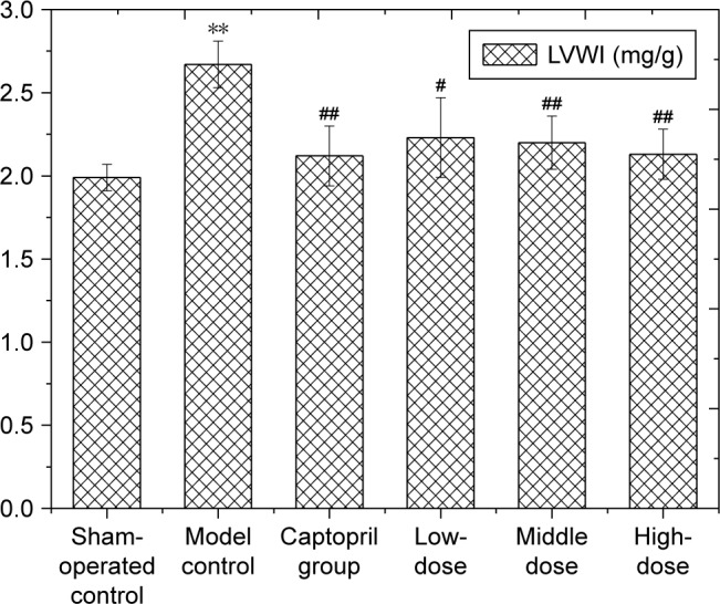
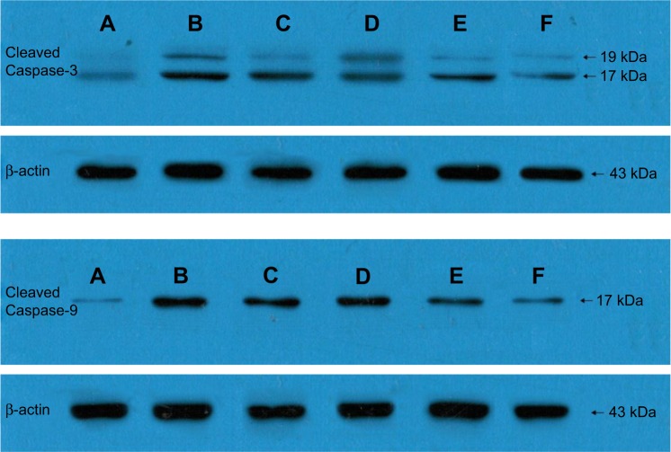
In this study, AMI model was established by ligating the anterior descending coronary artery in Wistar rats. After 4 weeks gavage of PF, the apparent signs and the left ventricle weight index of Wistar rats were observed. The left ventricular ejection fraction (LVEF) was evaluated by Doppler ultrasonography. Changes in cardiac morphology were observed by pathologic examination, and apoptosis was observed by the terminal deoxynucleotidyl transferase dUTP nick end labeling assay. In addition, enzyme-linked immunosorbent assay was used to detect the expression of tumor necrosis factor-α (TNF-α), interleukin-6 (IL-6) interleukin-10 (IL-10) and brain natriuretic peptide (BNP). Immunohistochemistry and Western blot method were applied to detect Caspase-3 and Caspase-9.
Compared with the model control, the survival conditions of rats in all treatment groups were generally improved after PF treatment. LVEF was significantly increased, and both left ventricular end-diastolic inner diameter and left ventricular end-systolic inner diameter were significantly reduced. Moreover, pathologic examination showed that the myocardium degeneration of the rats treated with PF was decreased, including neater arrangement, more complete myofilament, more uniform gap and less interstitial collagen fibers. Furthermore, the mitochondrial structure of cardiomyocytes was significantly improved. The ultrastructure was clear, and the arrangement of myofilament was more regular. Also, the expression of Caspase-3 and Caspase-9 was inhibited, and apoptosis was obviously reduced in the PF treatment groups. BNP, TNF-α and IL-6 were also decreased and IL-10 was increased in the treated rats.
PF could significantly improve the LVEF of rats. It decreased adverse left ventricular remodeling after myocardial infarction in rat models. The potential mechanism could be that PF decreased and inhibited BNP, TNF-α and IL-6, increased IL-10 and further inhibited the expression of Caspase-3 and Caspase-9, thus promoting ventricular remodeling.
芍药苷(PF)是芍药或牡丹的活性成分。因此,本研究旨在评估PF对急性心肌梗死(AMI)大鼠心室重构的改善作用及其机制。
本研究通过结扎Wistar大鼠冠状动脉前降支建立AMI模型。PF灌胃4周后,观察Wistar大鼠的明显体征及左心室重量指数。采用多普勒超声心动图评估左心室射血分数(LVEF)。通过病理检查观察心脏形态变化,采用末端脱氧核苷酸转移酶介导的dUTP缺口末端标记法观察细胞凋亡情况。此外,采用酶联免疫吸附测定法检测肿瘤坏死因子-α(TNF-α)、白细胞介素-6(IL-6)、白细胞介素-10(IL-10)和脑钠肽(BNP)的表达。应用免疫组织化学和蛋白质印迹法检测半胱天冬酶-3(Caspase-3)和半胱天冬酶-9(Caspase-9)。
与模型对照组相比,PF治疗后各治疗组大鼠的生存状况总体改善。LVEF显著升高,左心室舒张末期内径和左心室收缩末期内径均显著降低。此外,病理检查显示,PF治疗大鼠的心肌变性减轻,包括排列更整齐、肌丝更完整、间隙更均匀且间质胶原纤维更少。此外,心肌细胞的线粒体结构显著改善。超微结构清晰,肌丝排列更规则。而且,PF治疗组中Caspase-3和Caspase-9的表达受到抑制,细胞凋亡明显减少。治疗大鼠的BNP、TNF-α和IL-6也降低,而IL-10升高。
PF可显著提高大鼠的LVEF。它可减轻大鼠模型心肌梗死后不良的左心室重构。潜在机制可能是PF降低并抑制BNP、TNF-α和IL-6,升高IL-10,并进一步抑制Caspase-3和Caspase-9的表达,从而促进心室重构。