Hayashi Yuko, Miura Gen, Uzawa Akiyuki, Baba Takayuki, Yamamoto Shuichi
Department of Ophthalmology and Visual Science, Chiba University Graduate School of Medicine, Inohana 1-8-1, Chuo-ku, Chiba, 260-8670, Japan.
Department of Neurology, Graduate School of Medicine, Chiba University, Chiba, Japan.
BMC Neurol. 2018 Apr 25;18(1):52. doi: 10.1186/s12883-018-1051-2.
To present our findings in a case of convulsive seizures and loss of consciousness that developed during recording electroretinograms (ERG).
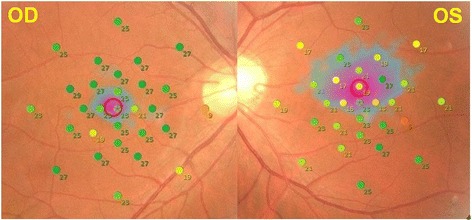
A 34-year-old man had reduced vision in his left eye for about 15 years, and night blindness for about two years. His visual acuity was 20/15 in the right eye and 20/50 in the left eye. The fundus was normal but the sensitivity in the macular region of the left eye was decreased. Optical coherence tomography (OCT) showed partial loss of the interdigitation zone. Upon completion of the flicker ERG recording, a paralysis developed in both upper limbs, then convulsions of the lower limbs followed by a loss of consciousness. The convulsions disappeared after an intravenous injection of diazepam. After that incident, he reported that he had had previous conscious-loss seizures.
Photosensitive epileptic seizures can occur with the light stimuli used for conventional ERG recordings. We recommended that clinicians request information on any prior seizure episodes of the patients and their family members before ERG recordings.
报告我们在记录视网膜电图(ERG)期间发生惊厥性癫痫发作和意识丧失病例中的发现。
一名34岁男性,左眼视力下降约15年,夜盲约2年。右眼视力为20/15,左眼视力为20/50。眼底正常,但左眼黄斑区敏感性降低。光学相干断层扫描(OCT)显示指状交叉区部分缺失。在完成闪烁ERG记录后,双上肢出现麻痹,随后下肢抽搐,继之意识丧失。静脉注射地西泮后抽搐消失。那次事件后,他报告曾有过意识丧失性癫痫发作。
传统ERG记录所使用的光刺激可引发光敏性癫痫发作。我们建议临床医生在进行ERG记录前询问患者及其家属既往任何癫痫发作史。