• 文献检索
  • 文档翻译
  • 深度研究
  • 学术资讯
  • Suppr Zotero 插件Zotero 插件
  • 邀请有礼
  • 套餐&价格
  • 历史记录
应用&插件
Suppr Zotero 插件Zotero 插件浏览器插件Mac 客户端Windows 客户端微信小程序
定价
高级版会员购买积分包购买API积分包
服务
文献检索文档翻译深度研究API 文档MCP 服务
关于我们
关于 Suppr公司介绍联系我们用户协议隐私条款
关注我们

Suppr 超能文献

核心技术专利:CN118964589B侵权必究
粤ICP备2023148730 号-1Suppr @ 2026

文献检索

告别复杂PubMed语法,用中文像聊天一样搜索,搜遍4000万医学文献。AI智能推荐,让科研检索更轻松。

立即免费搜索

文件翻译

保留排版,准确专业,支持PDF/Word/PPT等文件格式,支持 12+语言互译。

免费翻译文档

深度研究

AI帮你快速写综述,25分钟生成高质量综述,智能提取关键信息,辅助科研写作。

立即免费体验

乳腺MRI影像组学:计算机提取与人工提取影像表型的比较

Breast MRI radiomics: comparison of computer- and human-extracted imaging phenotypes.

作者信息

Sutton Elizabeth J, Huang Erich P, Drukker Karen, Burnside Elizabeth S, Li Hui, Net Jose M, Rao Arvind, Whitman Gary J, Zuley Margarita, Ganott Marie, Bonaccio Ermelinda, Giger Maryellen L, Morris Elizabeth A

机构信息

1Department of Radiology, Memorial Sloan Kettering Cancer Center, 1275 York Ave, New York, NY 10065 USA.

2Division of Cancer Treatment and Diagnosis, National Cancer Institute, National Institutes of Health, 9609 Medical Center Drive, Rockville, MD 20892 USA.

出版信息

Eur Radiol Exp. 2017;1(1):22. doi: 10.1186/s41747-017-0025-2. Epub 2017 Nov 21.

DOI:10.1186/s41747-017-0025-2
PMID:29708200
原文链接:https://pmc.ncbi.nlm.nih.gov/articles/PMC5909355/
Abstract

BACKGROUND

In this study, we sought to investigate if computer-extracted magnetic resonance imaging (MRI) phenotypes of breast cancer could replicate human-extracted size and Breast Imaging-Reporting and Data System (BI-RADS) imaging phenotypes using MRI data from The Cancer Genome Atlas (TCGA) project of the National Cancer Institute.

METHODS

Our retrospective interpretation study involved analysis of Health Insurance Portability and Accountability Act-compliant breast MRI data from The Cancer Imaging Archive, an open-source database from the TCGA project. This study was exempt from institutional review board approval at Memorial Sloan Kettering Cancer Center and the need for informed consent was waived. Ninety-one pre-operative breast MRIs with verified invasive breast cancers were analysed. Three fellowship-trained breast radiologists evaluated the index cancer in each case according to size and the BI-RADS lexicon for shape, margin, and enhancement (human-extracted image phenotypes [HEIP]). Human inter-observer agreement was analysed by the intra-class correlation coefficient (ICC) for size and Krippendorff's α for other measurements. Quantitative MRI radiomics of computerised three-dimensional segmentations of each cancer generated computer-extracted image phenotypes (CEIP). Spearman's rank correlation coefficients were used to compare HEIP and CEIP.

RESULTS

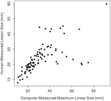
Inter-observer agreement for HEIP varied, with the highest agreement seen for size (ICC 0.679) and shape (ICC 0.527). The computer-extracted maximum linear size replicated the human measurement with  < 10. CEIP of shape, specifically sphericity and irregularity, replicated HEIP with both values < 0.001. CEIP did not demonstrate agreement with HEIP of tumour margin or internal enhancement.

CONCLUSIONS

Quantitative radiomics of breast cancer may replicate human-extracted tumour size and BI-RADS imaging phenotypes, thus enabling precision medicine.

摘要

背景

在本研究中,我们试图利用美国国立癌症研究所癌症基因组图谱(TCGA)项目的磁共振成像(MRI)数据,调查计算机提取的乳腺癌MRI表型是否能够复制人工提取的大小及乳腺影像报告和数据系统(BI-RADS)成像表型。

方法

我们的回顾性解读研究涉及对来自癌症影像存档库(TCGA项目的一个开源数据库)的符合《健康保险流通与责任法案》的乳腺MRI数据进行分析。本研究在纪念斯隆凯特琳癌症中心无需机构审查委员会批准,且无需获得知情同意。对91例术前经证实的浸润性乳腺癌的乳腺MRI进行了分析。三名经过专科培训的乳腺放射科医生根据大小以及BI-RADS词典中关于形态、边缘和强化的内容对每例中的索引癌进行评估(人工提取的图像表型[HEIP])。通过组内相关系数(ICC)分析大小方面的观察者间一致性,通过Krippendorff's α分析其他测量指标的观察者间一致性。对每种癌症的计算机三维分割进行定量MRI放射组学分析,生成计算机提取的图像表型(CEIP)。使用Spearman等级相关系数比较HEIP和CEIP。

结果

HEIP的观察者间一致性各不相同,在大小方面一致性最高(ICC 0.679),在形态方面一致性较高(ICC 0.527)。计算机提取的最大线性大小与人工测量值的复制程度< 10。形态方面的CEIP,特别是球形度和不规则度,与HEIP的复制程度均< 0.001。CEIP与肿瘤边缘或内部强化的HEIP未显示出一致性。

结论

乳腺癌的定量放射组学可能复制人工提取的肿瘤大小和BI-RADS成像表型,从而实现精准医疗。

https://cdn.ncbi.nlm.nih.gov/pmc/blobs/5a42/6091697/02568e7ad3c8/41747_2017_25_Fig2_HTML.jpg

相似文献

1
Breast MRI radiomics: comparison of computer- and human-extracted imaging phenotypes.乳腺MRI影像组学:计算机提取与人工提取影像表型的比较
Eur Radiol Exp. 2017;1(1):22. doi: 10.1186/s41747-017-0025-2. Epub 2017 Nov 21.
2
Breast imaging reporting and data system (BI-RADS) lexicon for breast MRI: interobserver variability in the description and assignment of BI-RADS category.乳腺磁共振成像的乳腺影像报告和数据系统(BI-RADS)词典:BI-RADS分类描述与指定中的观察者间差异
Eur J Radiol. 2015 Jan;84(1):71-76. doi: 10.1016/j.ejrad.2014.10.003. Epub 2014 Oct 16.
3
Breast lesion shape and margin evaluation: BI-RADS based metrics understate radiologists' actual levels of agreement.乳腺病变形状和边界评估:BI-RADS 度量标准低估了放射科医生的实际一致性水平。
Comput Biol Med. 2018 May 1;96:294-298. doi: 10.1016/j.compbiomed.2018.04.005. Epub 2018 Apr 13.
4
MR Imaging Radiomics Signatures for Predicting the Risk of Breast Cancer Recurrence as Given by Research Versions of MammaPrint, Oncotype DX, and PAM50 Gene Assays.用于预测MammaPrint、Oncotype DX和PAM50基因检测研究版本所给出的乳腺癌复发风险的磁共振成像放射组学特征
Radiology. 2016 Nov;281(2):382-391. doi: 10.1148/radiol.2016152110. Epub 2016 May 5.
5
Inter- and intra-observer agreement of BI-RADS-based subjective visual estimation of amount of fibroglandular breast tissue with magnetic resonance imaging: comparison to automated quantitative assessment.基于BI-RADS的乳腺纤维腺组织量磁共振成像主观视觉评估的观察者间及观察者内一致性:与自动定量评估的比较
Eur Radiol. 2016 Nov;26(11):3917-3922. doi: 10.1007/s00330-016-4274-x. Epub 2016 Apr 23.
6
Relationships Between MRI Breast Imaging-Reporting and Data System (BI-RADS) Lexicon Descriptors and Breast Cancer Molecular Subtypes: Internal Enhancement is Associated with Luminal B Subtype.MRI乳腺影像报告和数据系统(BI-RADS)术语描述符与乳腺癌分子亚型之间的关系:内部强化与管腔B亚型相关。
Breast J. 2017 Sep;23(5):579-582. doi: 10.1111/tbj.12799. Epub 2017 Mar 13.
7
Use of BI-RADS-MRI descriptors for differentiation between mucinous carcinoma and fibroadenoma.使用乳腺影像报告和数据系统(BI-RADS)-MRI描述符鉴别黏液性癌和纤维腺瘤。
Eur J Radiol. 2016 Jun;85(6):1092-8. doi: 10.1016/j.ejrad.2016.03.012. Epub 2016 Mar 17.
8
Breast cancer MRI radiomics: An overview of algorithmic features and impact of inter-reader variability in annotating tumors.乳腺癌 MRI 放射组学:算法特征概述及在肿瘤标注中读者间变异性的影响。
Med Phys. 2018 Jul;45(7):3076-3085. doi: 10.1002/mp.12925. Epub 2018 May 11.
9
Improved characterization of sub-centimeter enhancing breast masses on MRI with radiomics and machine learning in BRCA mutation carriers.利用放射组学和机器学习技术对 BRCA 基因突变携带者的亚厘米强化乳腺肿块进行改良特征描述。
Eur Radiol. 2020 Dec;30(12):6721-6731. doi: 10.1007/s00330-020-06991-7. Epub 2020 Jun 27.
10
Relationships Between Human-Extracted MRI Tumor Phenotypes of Breast Cancer and Clinical Prognostic Indicators Including Receptor Status and Molecular Subtype.人类提取的乳腺癌MRI肿瘤表型与包括受体状态和分子亚型在内的临床预后指标之间的关系。
Curr Probl Diagn Radiol. 2019 Sep-Oct;48(5):467-472. doi: 10.1067/j.cpradiol.2018.08.003. Epub 2018 Aug 23.

引用本文的文献

1
Heterogeneity Assessment of Breast Cancer Tumor Microenvironment: Multiparametric Quantitative Analysis with DCE-MRI and Discovery of Radiomics Biomarkers.乳腺癌肿瘤微环境的异质性评估:基于动态对比增强磁共振成像的多参数定量分析及影像组学生物标志物的发现
Breast Cancer (Dove Med Press). 2025 Jul 8;17:573-581. doi: 10.2147/BCTT.S530834. eCollection 2025.
2
Lymph node metastasis prediction and biological pathway associations underlying DCE-MRI deep learning radiomics in invasive breast cancer.基于深度学习的 MRI 放射组学预测浸润性乳腺癌淋巴结转移和生物学通路分析
BMC Med Imaging. 2024 Apr 16;24(1):91. doi: 10.1186/s12880-024-01255-y.
3

本文引用的文献

1
Quantitative MRI radiomics in the prediction of molecular classifications of breast cancer subtypes in the TCGA/TCIA data set.在TCGA/TCIA数据集中,定量MRI影像组学对乳腺癌亚型分子分类的预测
NPJ Breast Cancer. 2016;2:16012-. doi: 10.1038/npjbcancer.2016.12. Epub 2016 May 11.
2
MR Imaging Radiomics Signatures for Predicting the Risk of Breast Cancer Recurrence as Given by Research Versions of MammaPrint, Oncotype DX, and PAM50 Gene Assays.用于预测MammaPrint、Oncotype DX和PAM50基因检测研究版本所给出的乳腺癌复发风险的磁共振成像放射组学特征
Radiology. 2016 Nov;281(2):382-391. doi: 10.1148/radiol.2016152110. Epub 2016 May 5.
3
A quantitative heterogeneity analysis approach to molecular subtype recognition of breast cancer in dynamic contrast-enhanced magnetic imaging images from radiomics data.
一种基于放射组学数据的动态对比增强磁共振成像图像中乳腺癌分子亚型识别的定量异质性分析方法。
Quant Imaging Med Surg. 2023 Jul 1;13(7):4429-4446. doi: 10.21037/qims-22-1230. Epub 2023 May 26.
4
Radiomics model to classify mammary masses using breast DCE-MRI compared to the BI-RADS classification performance.使用乳腺动态对比增强磁共振成像(DCE-MRI)的放射组学模型对乳腺肿块进行分类,并与乳腺影像报告和数据系统(BI-RADS)分类性能进行比较。
Insights Imaging. 2023 Apr 13;14(1):64. doi: 10.1186/s13244-023-01404-x.
5
Applying artificial intelligence technology to assist with breast cancer diagnosis and prognosis prediction.应用人工智能技术辅助乳腺癌诊断及预后预测。
Front Oncol. 2022 Aug 31;12:980793. doi: 10.3389/fonc.2022.980793. eCollection 2022.
6
Effect of biopsy on the MRI radiomics classification of benign lesions and luminal A cancers.活检对良性病变和管腔A型癌的MRI影像组学分类的影响。
J Med Imaging (Bellingham). 2019 Jul;6(3):031408. doi: 10.1117/1.JMI.6.3.031408. Epub 2019 Feb 18.
7
Breast Lesion Classification with Multiparametric Breast MRI Using Radiomics and Machine Learning: A Comparison with Radiologists' Performance.使用影像组学和机器学习的多参数乳腺MRI进行乳腺病变分类:与放射科医生表现的比较
Cancers (Basel). 2022 Mar 29;14(7):1743. doi: 10.3390/cancers14071743.
8
Radiomics feature reliability assessed by intraclass correlation coefficient: a systematic review.通过组内相关系数评估的影像组学特征可靠性:一项系统评价
Quant Imaging Med Surg. 2021 Oct;11(10):4431-4460. doi: 10.21037/qims-21-86.
9
Differentiating Breast Tumors from Background Parenchymal Enhancement at Contrast-Enhanced Mammography: The Role of Radiomics-A Pilot Reader Study.在对比增强乳腺钼靶摄影中区分乳腺肿瘤与背景实质强化:影像组学的作用——一项初步阅片者研究
Diagnostics (Basel). 2021 Jul 13;11(7):1248. doi: 10.3390/diagnostics11071248.
10
Machine learning analysis of TCGA cancer data.TCGA癌症数据的机器学习分析。
PeerJ Comput Sci. 2021 Jul 12;7:e584. doi: 10.7717/peerj-cs.584. eCollection 2021.
Prediction of clinical phenotypes in invasive breast carcinomas from the integration of radiomics and genomics data.
通过整合放射组学和基因组学数据预测浸润性乳腺癌的临床表型
J Med Imaging (Bellingham). 2015 Oct;2(4):041007. doi: 10.1117/1.JMI.2.4.041007. Epub 2015 Sep 23.
4
Breast cancer molecular subtype classifier that incorporates MRI features.结合MRI特征的乳腺癌分子亚型分类器。
J Magn Reson Imaging. 2016 Jul;44(1):122-9. doi: 10.1002/jmri.25119. Epub 2016 Jan 12.
5
Breast MRI radiogenomics: Current status and research implications.乳腺MRI放射基因组学:现状与研究意义。
J Magn Reson Imaging. 2016 Jun;43(6):1269-78. doi: 10.1002/jmri.25116. Epub 2015 Dec 10.
6
Deciphering Genomic Underpinnings of Quantitative MRI-based Radiomic Phenotypes of Invasive Breast Carcinoma.解析浸润性乳腺癌基于定量MRI的放射组学表型的基因组基础
Sci Rep. 2015 Dec 7;5:17787. doi: 10.1038/srep17787.
7
Using computer-extracted image phenotypes from tumors on breast magnetic resonance imaging to predict breast cancer pathologic stage.利用乳腺磁共振成像上肿瘤的计算机提取图像表型预测乳腺癌病理分期。
Cancer. 2016 Mar 1;122(5):748-57. doi: 10.1002/cncr.29791. Epub 2015 Nov 30.
8
Radiogenomics: what it is and why it is important.放射基因组学:是什么以及为何重要。
J Am Coll Radiol. 2015 Aug;12(8):862-6. doi: 10.1016/j.jacr.2015.04.019.
9
Predicting classifier performance with limited training data: applications to computer-aided diagnosis in breast and prostate cancer.利用有限训练数据预测分类器性能:在乳腺癌和前列腺癌计算机辅助诊断中的应用
PLoS One. 2015 May 18;10(5):e0117900. doi: 10.1371/journal.pone.0117900. eCollection 2015.
10
Breast cancer subtype intertumor heterogeneity: MRI-based features predict results of a genomic assay.乳腺癌亚型间肿瘤异质性:基于MRI的特征可预测基因组检测结果。
J Magn Reson Imaging. 2015 Nov;42(5):1398-406. doi: 10.1002/jmri.24890. Epub 2015 Apr 7.