Muenzel Daniela, Daerr Heiner, Proksa Roland, Fingerle Alexander A, Kopp Felix K, Douek Philippe, Herzen Julia, Pfeiffer Franz, Rummeny Ernst J, Noël Peter B
1Department of Diagnostic and Interventional Radiology, Klinikum rechts der Isar, Technical University of Munich, Ismaningerstrasse 22, 81675 München, Germany.
2Philips GmbH Innovative Technologies, Research Laboratories, Hamburg, Germany.
Eur Radiol Exp. 2017;1(1):25. doi: 10.1186/s41747-017-0030-5. Epub 2017 Dec 22.
To assess the feasibility of dual-contrast spectral photon-counting computed tomography (SPCCT) for liver imaging.
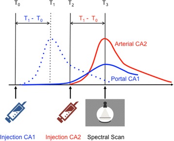
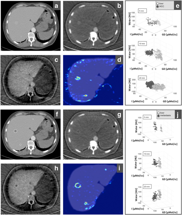
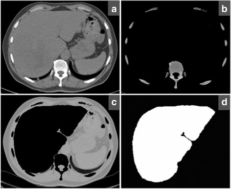
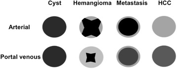
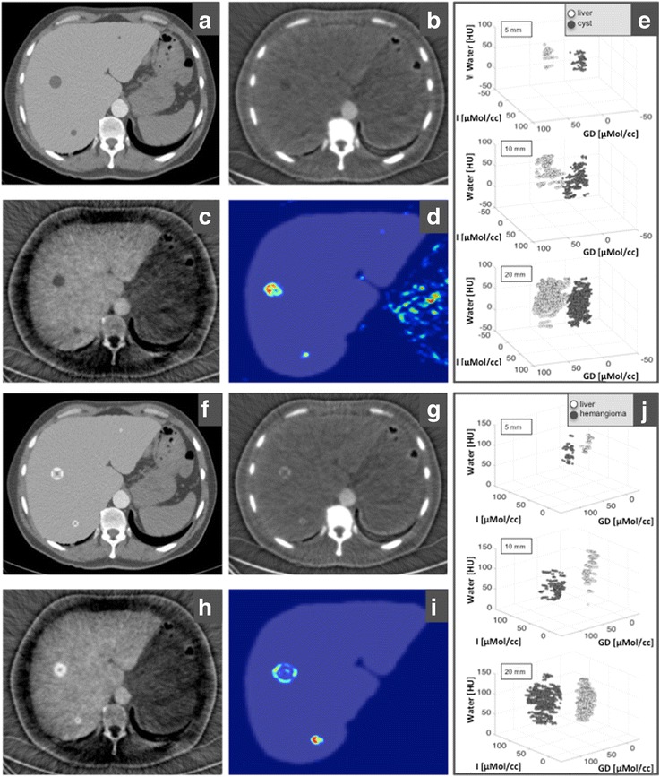
We present an SPCCT in-silico study for simultaneous mapping of the complementary distribution in the liver of two contrast agents (CAs) subsequently intravenously injected: a gadolinium-based contrast agent and an iodine-based contrast agent. Four types of simulated liver lesions with a characteristic arterial and portal venous pattern (haemangioma, hepatocellular carcinoma, cyst, and metastasis) are presented. A material decomposition was performed to reconstruct quantitative iodine and gadolinium maps. Finally, a multi-dimensional classification algorithm for automatic lesion detection is presented.
Our simulations showed that with a single-scan SPCCT and an adapted contrast injection protocol, it was possible to reconstruct contrast-enhanced images of the liver with arterial distribution of the iodine-based CA and portal venous phase of the gadolinium-based CA. The characteristic patterns of contrast enhancement were visible in all liver lesions. The approach allowed for an automatic detection and classification of liver lesions using a multi-dimensional analysis.
Dual-contrast SPCCT should be able to visualise the characteristic arterial and portal venous enhancement with a single scan, allowing for an automatic lesion detection and characterisation, with a reduced radiation exposure.
评估双能谱光子计数计算机断层扫描(SPCCT)用于肝脏成像的可行性。
我们开展了一项SPCCT计算机模拟研究,用于同时绘制两种随后经静脉注射的造影剂(CAs)在肝脏中的互补分布:一种基于钆的造影剂和一种基于碘的造影剂。展示了具有特征性动脉期和门静脉期模式的四种类型的模拟肝脏病变(血管瘤、肝细胞癌、囊肿和转移瘤)。进行了物质分解以重建定量碘图和钆图。最后,提出了一种用于自动病变检测的多维分类算法。
我们的模拟表明,通过单次扫描SPCCT和适配的造影剂注射方案,有可能重建出基于碘的造影剂动脉期分布和基于钆的造影剂门静脉期的肝脏对比增强图像。所有肝脏病变中均可见特征性的对比增强模式。该方法允许使用多维分析对肝脏病变进行自动检测和分类。
双能谱SPCCT应该能够通过单次扫描可视化特征性的动脉期和门静脉期增强,实现自动病变检测和特征描述,同时减少辐射暴露。