Zabihzadeh M, Moshirian T, Ghorbani M, Knaup C, Behrooz M A
Department of Medical Physics, Faculty of Medicine, Ahvaz Jundishapur University of Medical Sciences, Ahvaz, Iran.
Departments of Clinical Oncology, Golestan Hospital, Ahvaz Jundishapur University of Medical Sciences, Ahvaz, Iran.
J Biomed Phys Eng. 2018 Mar 1;8(1):13-28. eCollection 2018 Mar.
To enhance the dose to tumor, the use of high atomic number elements has been proposed.
The aim of this study is to investigate the effect of gold nanoparticle distribution on dose enhancement in tumor when the tumor is irradiated by typical monoenergetic X-ray beams by considering homogeneous and inhomogeneous distributions of gold nanoparticles (GNPs) in the tumor.
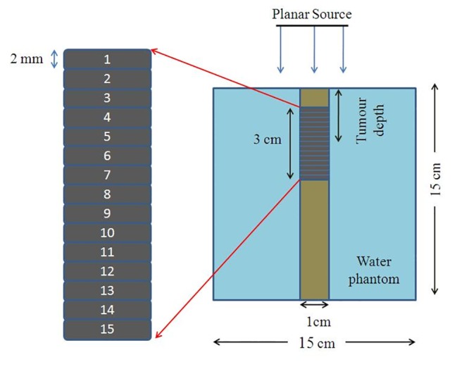
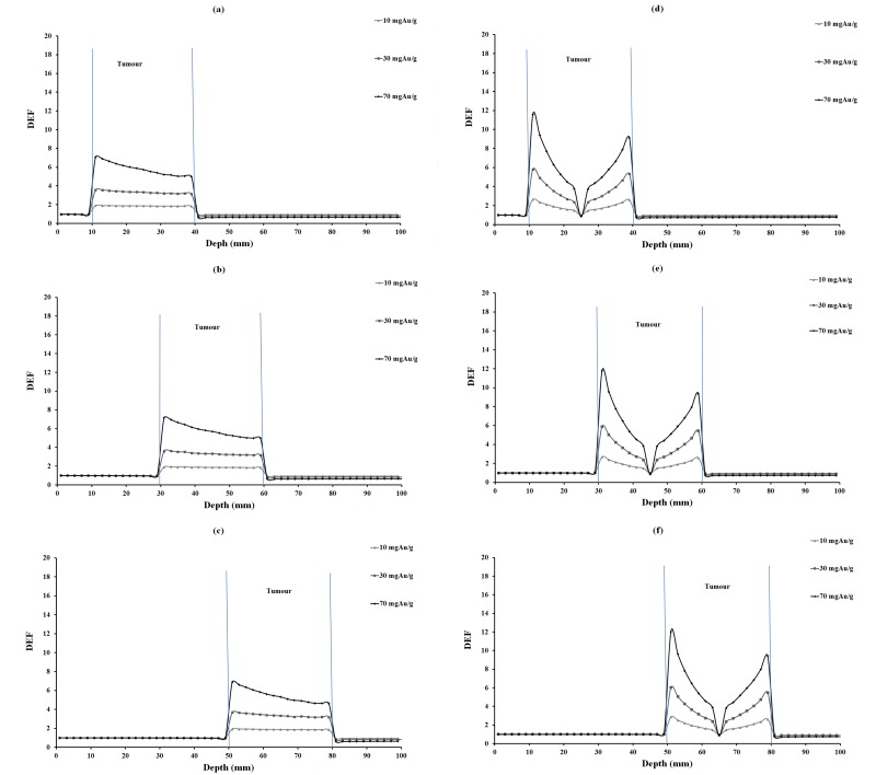
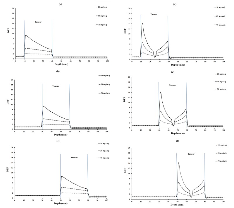
MCNP-4C Monte Carlo code was utilized for the simulation of a source, a phantom containing tumor and gold nanoparticles with concentrations of 10, 30 and 70 mg Au/g tumor. A 15 cm×15 cm×15 cm cubic water phantom was irradiated with a small planar source with four monoenergetic X-ray beams of 35, 55, 75 and 95 keV energy. Furthermore, tumor depths of 2.5 cm, 4.5 cm and 6.5 cm with homogeneous and inhomogeneous distributions of nanoparticles were studied. Each concentration, photon energy, tumor depth and type of distribution was evaluated in a separate simulation.
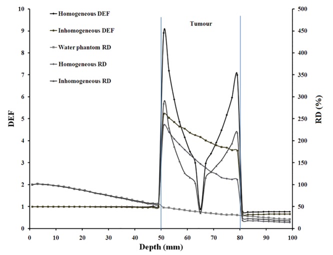
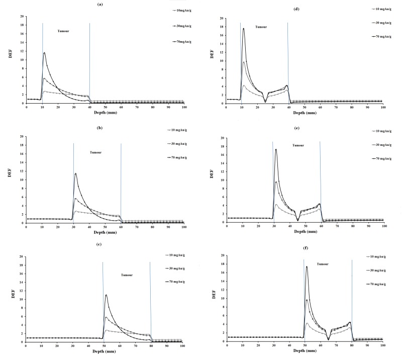
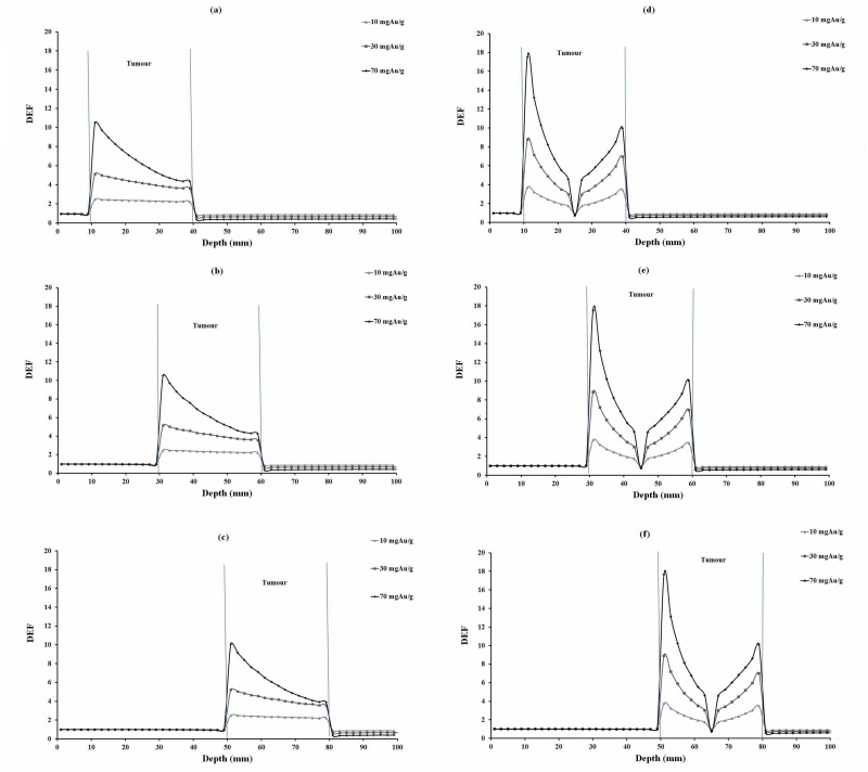
Results have shown that dose enhancement factor (DEF) in tumor increases approximately linearly with the concentration of gold nanoparticles. While DEF has fluctuations with photon energy, 55 keV photons have the highest DEF values compared to other energies. While DEF has relatively the same values with tumor located at various depths, inhomogeneous distribution of GNP has shown different results compared with the homogeneous model. Dose enhancement can be expected with relatively deep seated tumors in radiotherapy with low energy X-rays. Inhomogeneous model is recommended for the purpose of dose enhancement study because it mimics the real distribution of GNPs in tumor.
为提高肿瘤剂量,已有人提出使用高原子序数元素。
本研究旨在通过考虑金纳米颗粒(GNPs)在肿瘤中的均匀和非均匀分布,研究典型单能X射线束照射肿瘤时金纳米颗粒分布对肿瘤剂量增强的影响。
利用MCNP-4C蒙特卡罗代码模拟一个源、一个包含肿瘤和浓度分别为10、30和70 mg Au/g肿瘤的金纳米颗粒的体模。用一个小型平面源对一个15 cm×15 cm×15 cm的立方水体模进行照射,该平面源发出能量为35、55、75和95 keV的四束单能X射线。此外,还研究了纳米颗粒均匀和非均匀分布时肿瘤深度分别为2.5 cm、4.5 cm和6.5 cm的情况。在单独的模拟中对每种浓度、光子能量、肿瘤深度和分布类型进行评估。
结果表明,肿瘤中的剂量增强因子(DEF)随金纳米颗粒浓度近似线性增加。虽然DEF随光子能量有波动,但与其他能量相比,55 keV光子的DEF值最高。虽然DEF在不同深度的肿瘤处相对值相同,但GNP的非均匀分布与均匀模型相比显示出不同的结果。在低能X射线放疗中,对于位置相对较深的肿瘤可预期有剂量增强。由于非均匀模型模拟了GNPs在肿瘤中的实际分布,因此推荐用于剂量增强研究。