• 文献检索
  • 文档翻译
  • 深度研究
  • 学术资讯
  • Suppr Zotero 插件Zotero 插件
  • 邀请有礼
  • 套餐&价格
  • 历史记录
应用&插件
Suppr Zotero 插件Zotero 插件浏览器插件Mac 客户端Windows 客户端微信小程序
定价
高级版会员购买积分包购买API积分包
服务
文献检索文档翻译深度研究API 文档MCP 服务
关于我们
关于 Suppr公司介绍联系我们用户协议隐私条款
关注我们

Suppr 超能文献

核心技术专利:CN118964589B侵权必究
粤ICP备2023148730 号-1Suppr @ 2026

文献检索

告别复杂PubMed语法,用中文像聊天一样搜索,搜遍4000万医学文献。AI智能推荐,让科研检索更轻松。

立即免费搜索

文件翻译

保留排版,准确专业,支持PDF/Word/PPT等文件格式,支持 12+语言互译。

免费翻译文档

深度研究

AI帮你快速写综述,25分钟生成高质量综述,智能提取关键信息,辅助科研写作。

立即免费体验

一种用于皮肤镜图像中皮肤病变边界检测的新算法。

A New Algorithm for Skin Lesion Border Detection in Dermoscopy Images.

作者信息

Meskini E, Helfroush M S, Kazemi K, Sepaskhah M

机构信息

Department of Electrical and Electronics Engineering, Shiraz University of Technology, Shiraz, Iran.

Molecular Dermatology Research Center, Shiraz University of Medical Sciences, Shiraz, Iran.

出版信息

J Biomed Phys Eng. 2018 Mar 1;8(1):117-126. eCollection 2018 Mar.

PMID:29732346
原文链接:https://pmc.ncbi.nlm.nih.gov/articles/PMC5928301/
Abstract

BACKGROUND

With advances in medical imaging systems, digital dermoscopy has become one of the major imaging modalities in the analysis of skin lesions. Thus, automated segmentation or border detection has a great impact on the subsequent steps of skin cancer computer-aided diagnosis using demoscopy images. Since dermoscopy images suffer from artifacts such as shading and hair, there is a need for automated and robust artifact attenuation removal and lesion border detection.

METHODS

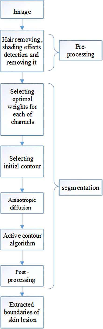
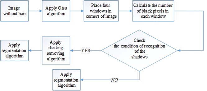
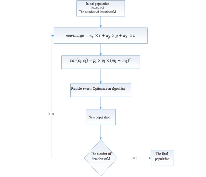
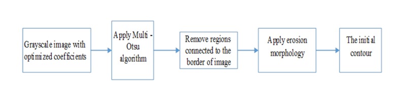
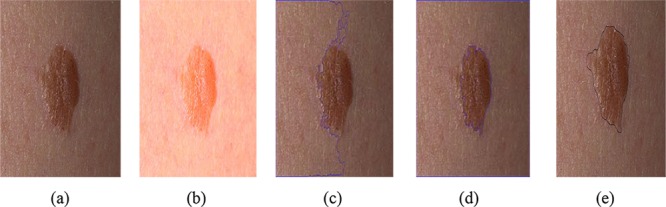
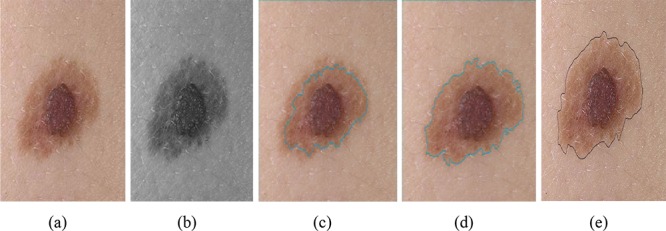
method for segmentation of dermoscopy images is proposed based on active contour. To this end, at first, a simple method for hair pixels is restored and a new scheme for shading detection is proposed. Then, particle swarm optimization (PSO) algorithm is applied to select the best coefficients for converting RGB to gray level. The obtained gray level image is then used as input for multi Otsu method which provides initial contour for border detection using active contour. Finally, Chan and Vese active contour is used for final lesion border detection.

RESULTS

The method is tested on a total of 145 dermoscopic images: 79 cases with benign lesion and 75 cases with melanoma lesion. Mean accuracy, sensitivity and specificity were obtained 94%, 78.5% and 99%, respectively.

CONCLUSION

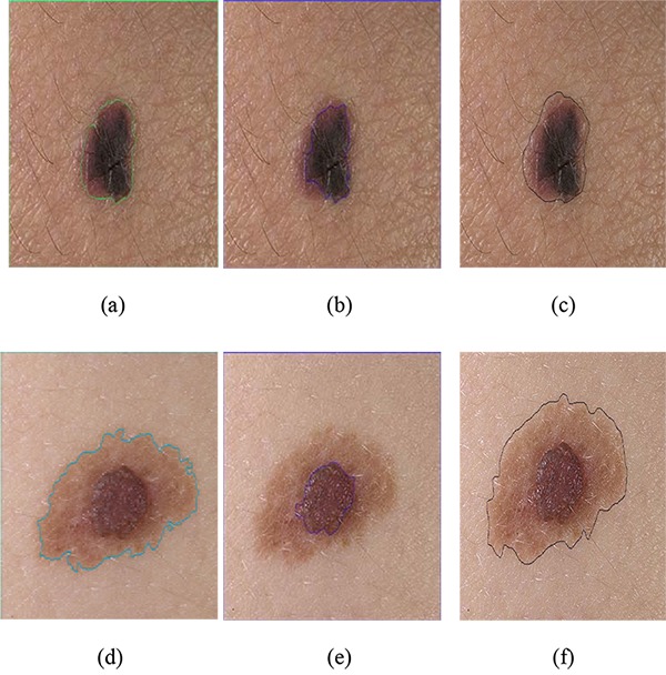
Results reveal that the proposed method segments the lesion from dermoscopy images accurately.

摘要

背景

随着医学成像系统的发展,数字皮肤镜检查已成为分析皮肤病变的主要成像方式之一。因此,自动分割或边界检测对使用皮肤镜图像的皮肤癌计算机辅助诊断的后续步骤有很大影响。由于皮肤镜图像存在诸如阴影和毛发等伪影,因此需要自动且强大的伪影衰减去除和病变边界检测。

方法

提出了一种基于活动轮廓的皮肤镜图像分割方法。为此,首先,恢复了一种简单的毛发像素处理方法,并提出了一种新的阴影检测方案。然后,应用粒子群优化(PSO)算法选择将RGB转换为灰度级的最佳系数。将获得的灰度图像用作多Otsu方法的输入,该方法为使用活动轮廓进行边界检测提供初始轮廓。最后,使用Chan和Vese活动轮廓进行最终的病变边界检测。

结果

该方法在总共145张皮肤镜图像上进行了测试:79例良性病变和75例黑色素瘤病变。平均准确率、灵敏度和特异性分别为94%、78.5%和99%。

结论

结果表明,所提出的方法能够准确地从皮肤镜图像中分割病变。

https://cdn.ncbi.nlm.nih.gov/pmc/blobs/c605/5928301/549cb80c8297/JBPE-8-117-g007.jpg
https://cdn.ncbi.nlm.nih.gov/pmc/blobs/c605/5928301/e53d3a36a995/JBPE-8-117-g001.jpg
https://cdn.ncbi.nlm.nih.gov/pmc/blobs/c605/5928301/51f977a328dd/JBPE-8-117-g002.jpg
https://cdn.ncbi.nlm.nih.gov/pmc/blobs/c605/5928301/abe641b75742/JBPE-8-117-g003.jpg
https://cdn.ncbi.nlm.nih.gov/pmc/blobs/c605/5928301/33a58e61ae07/JBPE-8-117-g004.jpg
https://cdn.ncbi.nlm.nih.gov/pmc/blobs/c605/5928301/dfcf325dcd6d/JBPE-8-117-g005.jpg
https://cdn.ncbi.nlm.nih.gov/pmc/blobs/c605/5928301/34e1a53d5769/JBPE-8-117-g006.jpg
https://cdn.ncbi.nlm.nih.gov/pmc/blobs/c605/5928301/549cb80c8297/JBPE-8-117-g007.jpg

相似文献

1
A New Algorithm for Skin Lesion Border Detection in Dermoscopy Images.一种用于皮肤镜图像中皮肤病变边界检测的新算法。
J Biomed Phys Eng. 2018 Mar 1;8(1):117-126. eCollection 2018 Mar.
2
Density-based parallel skin lesion border detection with webCL.基于密度的并行皮肤病变边界检测与WebCL技术
BMC Bioinformatics. 2015;16 Suppl 13(Suppl 13):S5. doi: 10.1186/1471-2105-16-S13-S5. Epub 2015 Sep 25.
3
Hair detection and lesion segmentation in dermoscopic images using domain knowledge.利用领域知识进行皮肤镜图像中的毛发检测和病灶分割。
Med Biol Eng Comput. 2018 Nov;56(11):2051-2065. doi: 10.1007/s11517-018-1837-9. Epub 2018 May 15.
4
Biologically inspired skin lesion segmentation using a geodesic active contour technique.使用测地线活动轮廓技术的生物启发式皮肤病变分割
Skin Res Technol. 2016 May;22(2):208-22. doi: 10.1111/srt.12252. Epub 2015 Sep 25.
5
Automatic skin tumour border detection for digital dermoscopy using a new digital image analysis scheme.使用一种新的数字图像分析方案对数字皮肤镜检查进行自动皮肤肿瘤边界检测。
Br J Biomed Sci. 2010;67(4):177-83. doi: 10.1080/09674845.2010.11730316.
6
An effective hair removal algorithm for dermoscopy images.一种有效的皮肤镜图像脱毛算法。
Skin Res Technol. 2013 Aug;19(3):230-5. doi: 10.1111/srt.12015. Epub 2013 Apr 7.
7
Lesion detection in demoscopy images with novel density-based and active contour approaches.基于密度和主动轮廓的新法检测皮肤镜图像中的损伤。
BMC Bioinformatics. 2010 Oct 7;11 Suppl 6(Suppl 6):S23. doi: 10.1186/1471-2105-11-S6-S23.
8
Automatic lesion border selection in dermoscopy images using morphology and color features.利用形态学和颜色特征自动选择皮肤镜图像中的病变边界。
Skin Res Technol. 2019 Jul;25(4):544-552. doi: 10.1111/srt.12685. Epub 2019 Mar 14.
9
A perceptually oriented method for contrast enhancement and segmentation of dermoscopy images.一种基于感知的皮肤镜图像对比度增强和分割方法。
Skin Res Technol. 2013 Feb;19(1):e490-7. doi: 10.1111/j.1600-0846.2012.00670.x. Epub 2012 Aug 13.
10
Abrupt skin lesion border cutoff measurement for malignancy detection in dermoscopy images.用于皮肤镜图像中恶性肿瘤检测的突然皮肤病变边界截断测量
BMC Bioinformatics. 2016 Oct 6;17(Suppl 13):367. doi: 10.1186/s12859-016-1221-4.

引用本文的文献

1
Automated Segmentation of Abnormal Tissues in Medical Images.医学图像中异常组织的自动分割
J Biomed Phys Eng. 2021 Aug 1;11(4):415-424. doi: 10.31661/jbpe.v0i0.958. eCollection 2021 Aug.

本文引用的文献

1
Model-based classification methods of global patterns in dermoscopic images.基于模型的皮肤镜图像全局模式分类方法。
IEEE Trans Med Imaging. 2014 May;33(5):1137-47. doi: 10.1109/TMI.2014.2305769.
2
An effective hair removal algorithm for dermoscopy images.一种有效的皮肤镜图像脱毛算法。
Skin Res Technol. 2013 Aug;19(3):230-5. doi: 10.1111/srt.12015. Epub 2013 Apr 7.
3
Lesion border detection in dermoscopy images using ensembles of thresholding methods.利用阈值方法集进行皮肤镜图像的病灶边界检测。
Skin Res Technol. 2013 Feb;19(1):e252-8. doi: 10.1111/j.1600-0846.2012.00636.x. Epub 2012 Jun 7.
4
Gradient vector flow with mean shift for skin lesion segmentation.基于梯度向量流和均值漂移的皮肤病变分割。
Comput Med Imaging Graph. 2011 Mar;35(2):121-7. doi: 10.1016/j.compmedimag.2010.08.002. Epub 2010 Sep 15.
5
Border detection in dermoscopy images using statistical region merging.使用统计区域合并的皮肤镜图像边界检测
Skin Res Technol. 2008 Aug;14(3):347-53. doi: 10.1111/j.1600-0846.2008.00301.x.
6
Active contours without edges.无边缘活动轮廓。
IEEE Trans Image Process. 2001;10(2):266-77. doi: 10.1109/83.902291.
7
Cancer statistics, 2006.2006年癌症统计数据。
CA Cancer J Clin. 2006 Mar-Apr;56(2):106-30. doi: 10.3322/canjclin.56.2.106.