Department of BioSciences, Rice University, Houston, TX, 77005, USA.
Department of Diagnostic and Biomedical Sciences, University of Texas Health Science Center at Houston, School of Dentistry, Houston, TX, 77054, USA.
Sci Rep. 2018 May 8;8(1):7262. doi: 10.1038/s41598-018-25435-3.
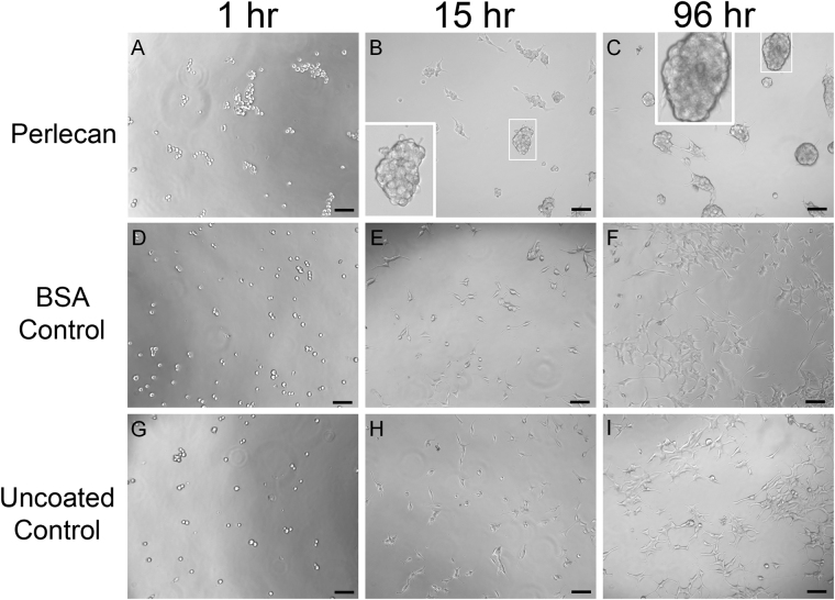
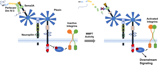
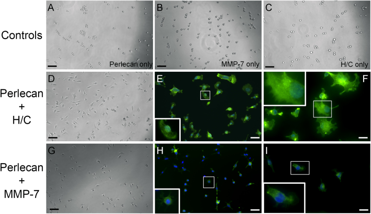
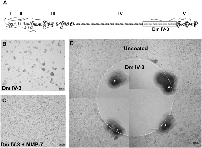
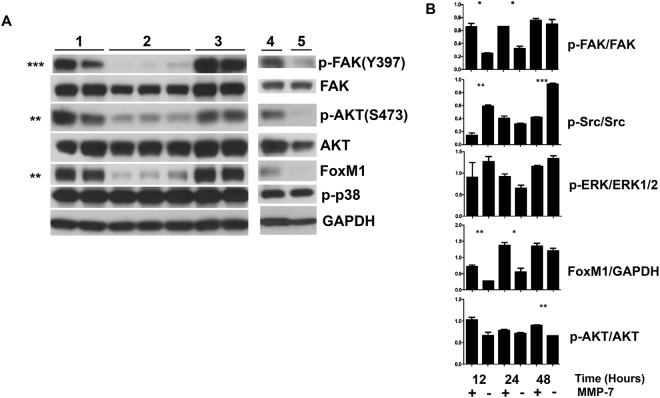
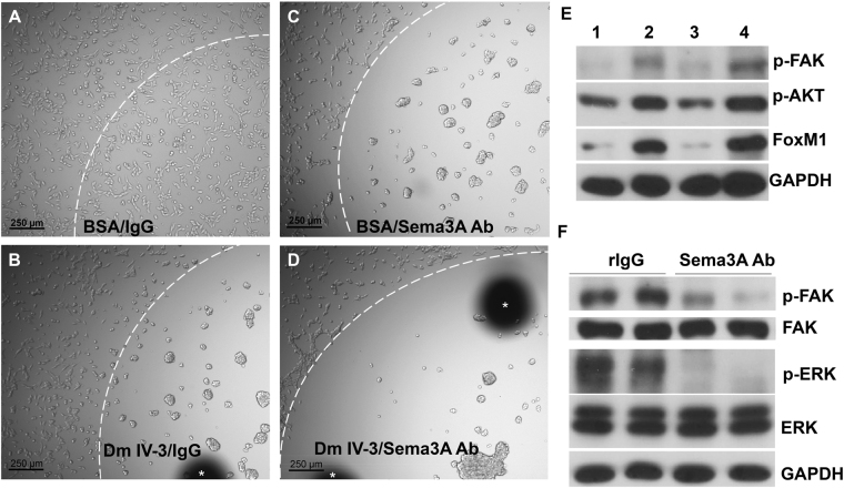
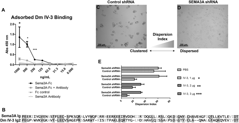
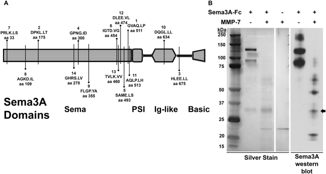
Interrupting the interplay between cancer cells and extracellular matrix (ECM) is a strategy to halt tumor progression and stromal invasion. Perlecan/heparan sulfate proteoglycan 2 (HSPG2) is an extracellular proteoglycan that orchestrates tumor angiogenesis, proliferation, differentiation and invasion. Metastatic prostate cancer (PCa) cells degrade perlecan-rich tissue borders to reach bone, including the basement membrane, vasculature, reactive stromal matrix and bone marrow. Domain IV-3, perlecan's last 7 immunoglobulin repeats, mimics native proteoglycan by promoting tumoroid formation. This is reversed by matrilysin/matrix metalloproteinase-7 (MMP-7) cleavage to favor cell dispersion and tumoroid dyscohesion. Both perlecan and Domain IV-3 induced a strong focal adhesion kinase (FAK) dephosphorylation/deactivation. MMP-7 cleavage of perlecan reversed this, with FAK in dispersed tumoroids becoming phosphorylated/activated with metastatic phenotype. We demonstrated Domain IV-3 interacts with the axon guidance protein semaphorin 3A (Sema3A) on PCa cells to deactivate pro-metastatic FAK. Sema3A antibody mimicked the Domain IV-3 clustering activity. Direct binding experiments showed Domain IV-3 binds Sema3A. Knockdown of Sema3A prevented Domain IV-3-induced tumoroid formation and Sema3A was sensitive to MMP-7 proteolysis. The perlecan-Sema3A complex abrogates FAK activity and stabilizes PCa cell interactions. MMP-7 expressing cells destroy the complex to initiate metastasis, destroy perlecan-rich borders, and favor invasion and progression to lethal bone disease.
中断癌细胞与细胞外基质(ECM)的相互作用是阻止肿瘤进展和基质浸润的一种策略。多配体蛋白聚糖 2(HSPG2)是一种细胞外蛋白聚糖,可协调肿瘤血管生成、增殖、分化和侵袭。转移性前列腺癌(PCa)细胞降解富含硫酸乙酰肝素蛋白聚糖的组织边界以到达骨骼,包括基底膜、脉管系统、反应性基质和骨髓。多配体蛋白聚糖的最后 7 个免疫球蛋白重复序列 IV-3 模拟天然蛋白聚糖,通过促进肿瘤球形成来促进肿瘤发生。基质金属蛋白酶-7(MMP-7)的切割将其逆转,有利于细胞分散和肿瘤球解聚。多配体蛋白聚糖和 IV-3 结构域都诱导强烈的粘着斑激酶(FAK)去磷酸化/失活。多配体蛋白聚糖的 MMP-7 切割逆转了这一点,分散的肿瘤球中的 FAK 发生磷酸化/激活,具有转移表型。我们证明 IV-3 结构域与 PCa 细胞上的轴突导向蛋白神经纤毛蛋白 3A(Sema3A)相互作用,使促转移的 FAK 失活。Sema3A 抗体模拟了 IV-3 结构域的聚集活性。直接结合实验表明 IV-3 结构域结合 Sema3A。Sema3A 的敲低阻止了 IV-3 结构域诱导的肿瘤球形成,Sema3A 对 MMP-7 蛋白水解敏感。多配体蛋白聚糖-Sema3A 复合物使 FAK 失活并稳定 PCa 细胞相互作用。表达 MMP-7 的细胞破坏复合物以启动转移,破坏富含多配体蛋白聚糖的边界,并有利于侵袭和进展为致命的骨疾病。