• 文献检索
  • 文档翻译
  • 深度研究
  • 学术资讯
  • Suppr Zotero 插件Zotero 插件
  • 邀请有礼
  • 套餐&价格
  • 历史记录
应用&插件
Suppr Zotero 插件Zotero 插件浏览器插件Mac 客户端Windows 客户端微信小程序
定价
高级版会员购买积分包购买API积分包
服务
文献检索文档翻译深度研究API 文档MCP 服务
关于我们
关于 Suppr公司介绍联系我们用户协议隐私条款
关注我们

Suppr 超能文献

核心技术专利:CN118964589B侵权必究
粤ICP备2023148730 号-1Suppr @ 2026

文献检索

告别复杂PubMed语法,用中文像聊天一样搜索,搜遍4000万医学文献。AI智能推荐,让科研检索更轻松。

立即免费搜索

文件翻译

保留排版,准确专业,支持PDF/Word/PPT等文件格式,支持 12+语言互译。

免费翻译文档

深度研究

AI帮你快速写综述,25分钟生成高质量综述,智能提取关键信息,辅助科研写作。

立即免费体验

应用弥散张量成像、静息态功能磁共振成像和磁敏感加权成像检测 Wilson 病患者的神经症状不对称性。

The asymmetry of neural symptoms in Wilson's disease patients detecting by diffusion tensor imaging, resting-state functional MRI, and susceptibility-weighted imaging.

机构信息

Department of Neurology The East Area of the First Affiliated Hospital Sun Yat-Sen University Guangzhou China.

Department of Neurology The First Affiliated Hospital Sun Yat-Sen University Guangzhou China.

出版信息

Brain Behav. 2018 Apr 14;8(5):e00930. doi: 10.1002/brb3.930. eCollection 2018 May.

DOI:10.1002/brb3.930
PMID:29761003
原文链接:https://pmc.ncbi.nlm.nih.gov/articles/PMC5943770/
Abstract

OBJECTIVE

To investigate the cause of the motor asymmetry in Wilson's disease (WD) patients using functional MRI.

METHODS

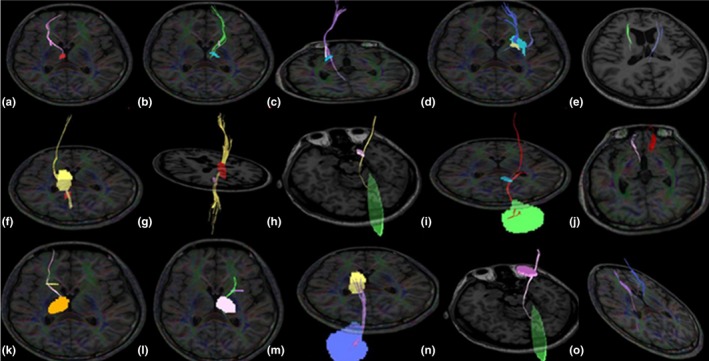
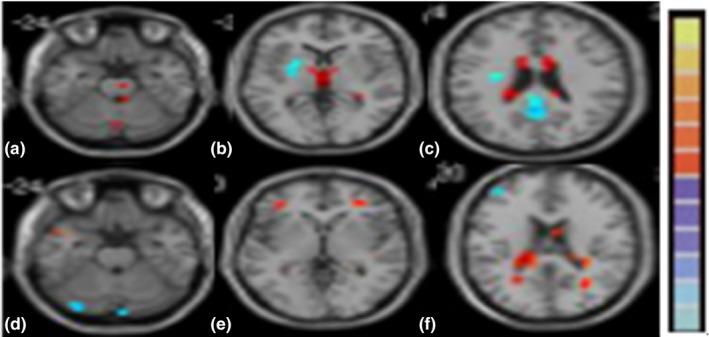
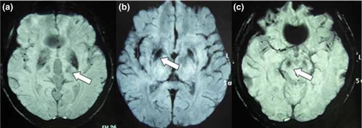
Fifty patients with WD and 20 age-matched healthy controls were enrolled. Neurological symptoms were scored using the modified Young Scale. All study subjects underwent diffusion tensor imaging (DTI), susceptibility-weighted imaging (SWI), and resting-state functional MRI (rs-fMRI) of the brain. Six regions of interest (ROI) were chosen. Fiber volumes between ROIs on DTI, corrected phase (CP) values on SWI, amplitude of low-frequency fluctuation (ALFF), and regional homogeneity (REHO) values on rs-fMRI were determined. Asymmetry index (right or left value/left or right value) was evaluated.

RESULTS

Asymmetry of rigidity, tremor, choreic movement, and gait abnormality (asymmetry index = 1.33, 1.39, 1.36, 1.40), fiber tracts between the GP and substantia nigra (SN), GP and PU, SN and thalamus (TH), SN and cerebellum, head of the caudate nucleus (CA) and SN, PU and CA, CA and TH, TH and cerebellum (asymmetry index = 1.233, 1.260, 1.269, 1.437, 1.503, 1.138, 1.145, 1.279), CP values in the TH, SN (asymmetry index = 1.327, 1.166), ALFF values, and REHO values of the TH (asymmetry index = 1.192, 1.233) were found. Positive correlation between asymmetry index of rigidity and fiber volumes between the GP and SN, SN and TH ( = .221, .133,  = .043, .036), and tremor and fiber volumes between the CA and TH ( = .045,  = .040) was found.

CONCLUSIONS

The neurological symptoms of patients with WD were asymmetry. The asymmetry of fiber projections may be the main cause of motor asymmetry in patients with WD.

摘要

目的

使用功能磁共振成像(fMRI)研究 Wilson 病(WD)患者运动不对称的原因。

方法

纳入 50 例 WD 患者和 20 例年龄匹配的健康对照者。使用改良 Young 量表对神经症状进行评分。所有研究对象均接受弥散张量成像(DTI)、磁敏感加权成像(SWI)和脑静息态功能磁共振(rs-fMRI)检查。选择 6 个感兴趣区(ROI)。确定 DTI 上 ROI 之间的纤维体积、SWI 上校正相位(CP)值、低频波动幅度(ALFF)和 rs-fMRI 上区域同质性(REHO)值。评估不对称指数(右侧或左侧值/左侧或右侧值)。

结果

僵硬、震颤、舞蹈运动和步态异常的不对称性(不对称指数=1.33、1.39、1.36、1.40),苍白球(GP)与黑质(SN)、GP 与壳核(PU)、SN 与丘脑(TH)、SN 与小脑、尾状核头部(CA)与 SN、PU 与 CA、CA 与 TH、TH 与小脑之间的纤维束(不对称指数=1.233、1.260、1.269、1.437、1.503、1.138、1.145、1.279),TH、SN 中的 CP 值(不对称指数=1.327、1.166)、ALFF 值和 TH 的 REHO 值(不对称指数=1.192、1.233)。僵硬的不对称指数与 GP 和 SN、SN 和 TH 之间的纤维体积呈正相关(r=0.221、0.133),震颤与 CA 和 TH 之间的纤维体积呈正相关(r=0.045、0.040)。

结论

WD 患者的神经症状为不对称性。纤维投射的不对称性可能是 WD 患者运动不对称的主要原因。

https://cdn.ncbi.nlm.nih.gov/pmc/blobs/62b6/5943770/e009e38df72d/BRB3-8-e00930-g003.jpg
https://cdn.ncbi.nlm.nih.gov/pmc/blobs/62b6/5943770/0c7a1a12ac65/BRB3-8-e00930-g001.jpg
https://cdn.ncbi.nlm.nih.gov/pmc/blobs/62b6/5943770/981e599f52f1/BRB3-8-e00930-g002.jpg
https://cdn.ncbi.nlm.nih.gov/pmc/blobs/62b6/5943770/e009e38df72d/BRB3-8-e00930-g003.jpg
https://cdn.ncbi.nlm.nih.gov/pmc/blobs/62b6/5943770/0c7a1a12ac65/BRB3-8-e00930-g001.jpg
https://cdn.ncbi.nlm.nih.gov/pmc/blobs/62b6/5943770/981e599f52f1/BRB3-8-e00930-g002.jpg
https://cdn.ncbi.nlm.nih.gov/pmc/blobs/62b6/5943770/e009e38df72d/BRB3-8-e00930-g003.jpg

相似文献

1
The asymmetry of neural symptoms in Wilson's disease patients detecting by diffusion tensor imaging, resting-state functional MRI, and susceptibility-weighted imaging.应用弥散张量成像、静息态功能磁共振成像和磁敏感加权成像检测 Wilson 病患者的神经症状不对称性。
Brain Behav. 2018 Apr 14;8(5):e00930. doi: 10.1002/brb3.930. eCollection 2018 May.
2
Diffusion tensor imaging of the extracorticospinal network in the brains of patients with Wilson disease.肝豆状核变性患者大脑皮质脊髓外网络的扩散张量成像
J Neurol Sci. 2016 Mar 15;362:292-8. doi: 10.1016/j.jns.2016.02.006. Epub 2016 Feb 4.
3
[Difference in imaging and metal metabolism between hepatic and cerebral type Wilson disease].肝型和脑型威尔逊病在影像学及金属代谢方面的差异
Zhonghua Yi Xue Za Zhi. 2017 Jan 17;97(3):176-181. doi: 10.3760/cma.j.issn.0376-2491.2017.03.004.
4
Study on Lesion Assessment of Cerebello-Thalamo-Cortical Network in Wilson's Disease with Diffusion Tensor Imaging.应用弥散张量成像研究肝豆状核变性患者小脑-丘脑-皮质网络损伤。
Neural Plast. 2017;2017:7323121. doi: 10.1155/2017/7323121. Epub 2017 Jul 11.
5
Regional morphometric abnormalities and clinical relevance in Wilson's disease.Wilson 病的区域性形态计量异常及其临床相关性。
Mov Disord. 2019 Apr;34(4):545-554. doi: 10.1002/mds.27641. Epub 2019 Feb 28.
6
Substantia Nigra Volume Dissociates Bradykinesia and Rigidity from Tremor in Parkinson's Disease: A 7 Tesla Imaging Study.黑质体积可将帕金森病的运动迟缓与僵硬与震颤分开:7 特斯拉成像研究。
J Parkinsons Dis. 2020;10(2):591-604. doi: 10.3233/JPD-191890.
7
Brain microstructural abnormalities in patients with Wilson's disease: A systematic review of diffusion tenor imaging studies.威尔逊病患者的脑微观结构异常:扩散张量成像研究的系统综述
Brain Imaging Behav. 2022 Dec;16(6):2809-2840. doi: 10.1007/s11682-022-00733-7. Epub 2022 Nov 3.
8
Microstructure changes in whiter matter relate to cognitive impairment in Wilson's disease.脑白质微观结构变化与肝豆状核变性患者的认知障碍有关。
Biosci Rep. 2019 Mar 15;39(3). doi: 10.1042/BSR20181651. Print 2019 Mar 29.
9
Sonographic detection of basal ganglia lesions in asymptomatic and symptomatic Wilson disease.超声检查对无症状和有症状肝豆状核变性患者基底节病变的检测
Neurology. 2005 May 24;64(10):1726-32. doi: 10.1212/01.WNL.0000161847.46465.B9.
10
Susceptibility-Weighted Imaging Manifestations in the Brain of Wilson's Disease Patients.肝豆状核变性患者脑部的磁敏感加权成像表现
PLoS One. 2015 Apr 27;10(4):e0125100. doi: 10.1371/journal.pone.0125100. eCollection 2015.

引用本文的文献

1
Assessment of sleep disturbance in patients with Wilson's disease.评估威尔逊病患者的睡眠障碍。
BMC Psychiatry. 2024 Mar 13;24(1):205. doi: 10.1186/s12888-024-05493-w.
2
Machine learning for detecting Wilson's disease by amplitude of low-frequency fluctuation.基于低频波动幅度的机器学习用于检测威尔逊氏病
Heliyon. 2023 Jul 7;9(7):e18087. doi: 10.1016/j.heliyon.2023.e18087. eCollection 2023 Jul.
3
Neurological-Type Wilson Disease: Epidemiology, Clinical Manifestations, Diagnosis, and Management.神经型威尔逊病:流行病学、临床表现、诊断与治疗

本文引用的文献

1
Diffusion tensor imaging of the extracorticospinal network in the brains of patients with Wilson disease.肝豆状核变性患者大脑皮质脊髓外网络的扩散张量成像
J Neurol Sci. 2016 Mar 15;362:292-8. doi: 10.1016/j.jns.2016.02.006. Epub 2016 Feb 4.
2
Ipsilateral deficits of dopaminergic neurotransmission in Parkinson's disease.帕金森病中多巴胺能神经传递的同侧缺陷。
Ann Clin Transl Neurol. 2015 Dec 5;3(1):21-6. doi: 10.1002/acn3.268. eCollection 2016 Jan.
3
Diffusion tensor imaging (DTI) and its clinical correlates in drug naïve Wilson's disease.
Cureus. 2023 Apr 26;15(4):e38170. doi: 10.7759/cureus.38170. eCollection 2023 Apr.
4
Altered microstructural pattern of the cortex and basal forebrain cholinergic system in wilson's disease: an automated fiber quantification tractography study.威尔逊病中皮质和基底前脑胆碱能系统的微观结构模式改变:一项自动纤维定量纤维束成像研究
Brain Imaging Behav. 2023 Apr;17(2):200-212. doi: 10.1007/s11682-022-00753-3. Epub 2023 Jan 24.
5
Disrupted topological organization of the motor execution network in Wilson's disease.威尔逊病中运动执行网络拓扑组织的破坏
Front Neurol. 2022 Nov 21;13:1029669. doi: 10.3389/fneur.2022.1029669. eCollection 2022.
6
Brain microstructural abnormalities in patients with Wilson's disease: A systematic review of diffusion tenor imaging studies.威尔逊病患者的脑微观结构异常:扩散张量成像研究的系统综述
Brain Imaging Behav. 2022 Dec;16(6):2809-2840. doi: 10.1007/s11682-022-00733-7. Epub 2022 Nov 3.
7
An Updated Overview of the Magnetic Resonance Imaging of Brain Iron in Movement Disorders.运动障碍性脑铁磁共振成像的最新综述。
Behav Neurol. 2022 Feb 24;2022:3972173. doi: 10.1155/2022/3972173. eCollection 2022.
8
Neurologic impairment in Wilson disease.威尔逊病中的神经功能损害。
Ann Transl Med. 2019 Apr;7(Suppl 2):S64. doi: 10.21037/atm.2019.02.43.
9
Clinical presentations of Wilson disease.威尔逊病的临床表现。
Ann Transl Med. 2019 Apr;7(Suppl 2):S60. doi: 10.21037/atm.2019.04.27.
未经药物治疗的威尔逊病的弥散张量成像(DTI)及其临床相关性。
Metab Brain Dis. 2013 Sep;28(3):455-62. doi: 10.1007/s11011-013-9407-1. Epub 2013 May 1.
4
Susceptibility weighted imaging in the evaluation of movement disorders.磁敏感加权成像在运动障碍评估中的应用。
Clin Radiol. 2013 Jun;68(6):e338-48. doi: 10.1016/j.crad.2012.12.003. Epub 2013 Mar 26.
5
Clinical MRI for iron detection in Parkinson's disease.帕金森病中铁的临床 MRI 检测。
Clin Imaging. 2013 Jul-Aug;37(4):631-6. doi: 10.1016/j.clinimag.2013.02.001. Epub 2013 Mar 20.
6
Topographical differences of brain iron deposition between progressive supranuclear palsy and parkinsonian variant multiple system atrophy.进行性核上性麻痹与帕金森病变异型多系统萎缩脑铁沉积的解剖部位差异。
J Neurol Sci. 2013 Feb 15;325(1-2):29-35. doi: 10.1016/j.jns.2012.11.009. Epub 2012 Dec 20.
7
Widespread cerebral cortical mineralization in Wilson's disease detected by susceptibility-weighted imaging.肝豆状核变性患者脑皮质广泛矿化的磁敏感加权成像表现
J Neurol Sci. 2012 Feb 15;313(1-2):54-6. doi: 10.1016/j.jns.2011.09.031. Epub 2011 Oct 20.
8
Diffusion tensor imaging and colored fractional anisotropy mapping of the ventralis intermedius nucleus of the thalamus.丘脑腹中间核的弥散张量成像及彩色各向异性分数映射。
Neurosurgery. 2011 Nov;69(5):1124-9; discussion 1129-30. doi: 10.1227/NEU.0b013e3182296a42.
9
Individual fiber anatomy of the subthalamic region revealed with diffusion tensor imaging: a concept to identify the deep brain stimulation target for tremor suppression.应用弥散张量成像显示的丘脑底核的个体纤维解剖结构:用于识别抑制震颤的深部脑刺激靶点的概念。
Neurosurgery. 2011 Apr;68(4):1069-75; discussion 1075-6. doi: 10.1227/NEU.0b013e31820a1a20.
10
Utility of axial and radial diffusivity from diffusion tensor MRI as markers of neurodegeneration in amyotrophic lateral sclerosis.弥散张量 MRI 的轴向和径向弥散度作为肌萎缩侧索硬化症神经退行性变的标志物的效用。
Brain Res. 2010 Aug 12;1348:156-64. doi: 10.1016/j.brainres.2010.05.067. Epub 2010 Jun 1.