Department of Pharmacology, Faculty of Pharmacy, University of Debrecen, Nagyerdei Krt 98, 4032 Debrecen, Hungary.
Department of Anatomy, Histology and Embriology, Faculty of Medicine, University of Debrecen, Nagyerdei Krt 98, 4032 Debrecen, Hungary.
Molecules. 2018 May 15;23(5):1184. doi: 10.3390/molecules23051184.
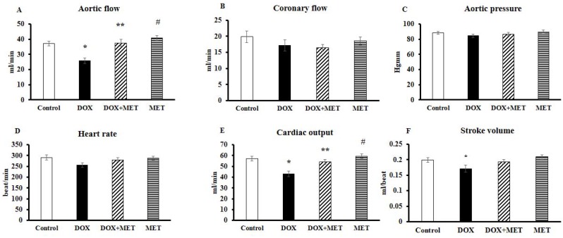
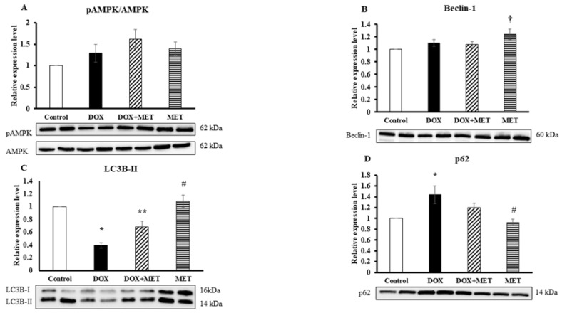
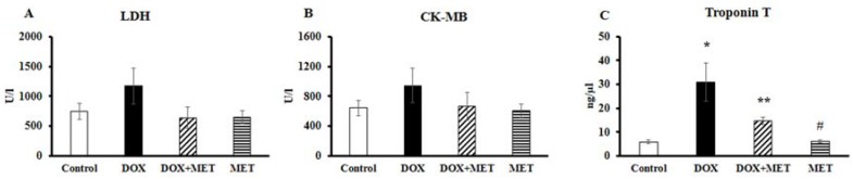
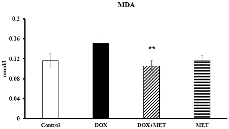
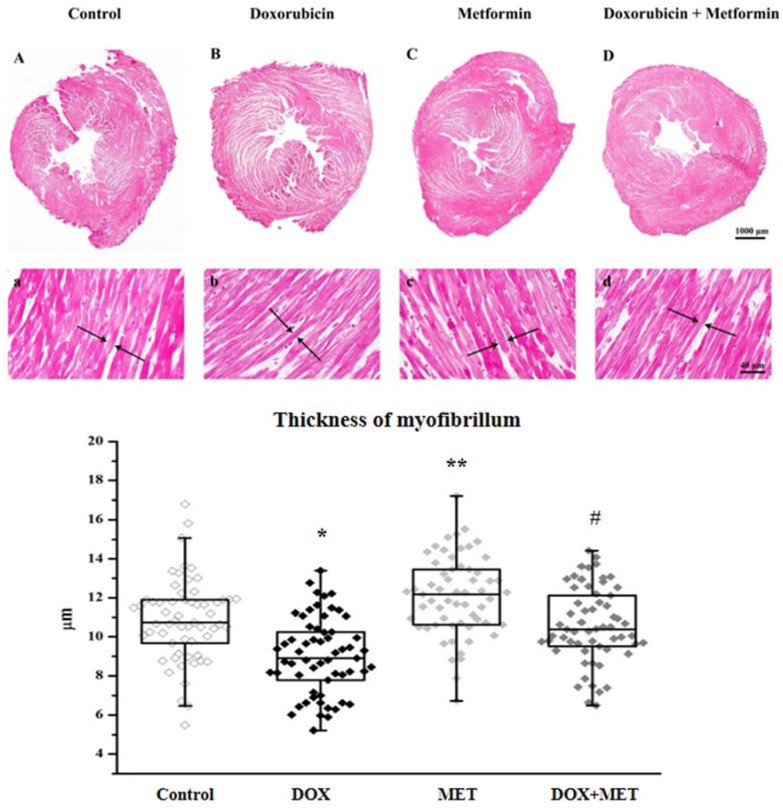
The molecular mechanisms underlying doxorubicin-induced cardiotoxicity are still being investigated, but are known to involve oxidative stress, mitochondrial dysfunction, and the dysregulation of autophagy. The objective of the current study was to examine the protective role of metformin and its effect on autophagy in doxorubicin-induced cardiotoxicity. Sprague⁻Dawley rats were divided into four groups at random. The doxorubicin-treated group received doxorubicin (3 mg/kg every second day) intraperitoneally. The metformin-treated group received 250 mg/kg/day metformin via gavage. The doxorubicin + metformin-treated group received both at the above-mentioned doses. The control group received vehicle only. Following the two-week treatment, the hearts were isolated, and cardiac functions were registered. Serum levels of lactate dehydrogenase (LDH), creatine kinase iso-enzyme MB (CK-MB) enzyme, Troponin T, and cardiac malondialdehyde (MDA) were also measured. Heart tissue samples were histopathologically examined by using Masson's trichrome staining and Western blot analysis was conducted for evaluating the expression level of AMP-activated protein kinase (AMPK) and autophagy-associated proteins beclin-1, LC3B-II, and p62, respectively. The results revealed that treatment with metformin conferred increased cardiac protection against the development of cardiotoxicity manifested by a significant decrease in serum Troponin T and cardiac MDA levels, and remarkable improvement in heart function in connection with histopathological features. Furthermore, by focusing on the contribution of autophagic proteins, it was found that metformin normalised autophagy, which may help cardiomyocytes survive doxorubicin-induced toxicity. These results promote the use of metformin, which would be a preferable drug for patients receiving doxorubicin.
多柔比星诱导心脏毒性的分子机制仍在研究中,但已知涉及氧化应激、线粒体功能障碍和自噬失调。本研究的目的是研究二甲双胍的保护作用及其对多柔比星诱导心脏毒性的自噬作用。将 Sprague-Dawley 大鼠随机分为四组。多柔比星处理组腹腔内给予多柔比星(每两天 3mg/kg)。二甲双胍处理组灌胃给予 250mg/kg/天二甲双胍。多柔比星+二甲双胍处理组给予上述两种药物。对照组仅给予载体。经过两周的治疗,分离心脏,记录心脏功能。还测量血清乳酸脱氢酶(LDH)、肌酸激酶同工酶 MB(CK-MB)酶、肌钙蛋白 T 和心脏丙二醛(MDA)水平。通过 Masson 三色染色对心脏组织样本进行组织病理学检查,并通过 Western blot 分析分别评估 AMP 激活的蛋白激酶(AMPK)和自噬相关蛋白 beclin-1、LC3B-II 和 p62 的表达水平。结果表明,二甲双胍治疗可显著提高心脏对心脏毒性的保护作用,表现为血清肌钙蛋白 T 和心脏 MDA 水平显著降低,与组织病理学特征相关的心功能显著改善。此外,通过关注自噬蛋白的贡献,发现二甲双胍可使自噬正常化,这可能有助于心肌细胞在多柔比星诱导的毒性中存活。这些结果促进了二甲双胍的使用,对于接受多柔比星治疗的患者来说,二甲双胍将是一种更优的药物。