University Children's Hospital, University of Wuerzburg, Josef-Schneider-Str. 2, 97080, Wuerzburg, Germany.
Department of Neurology, University of Wuerzburg, Josef-Schneider-Str. 11, 97080, Wuerzburg, Germany.
J Neuroinflammation. 2018 May 23;15(1):156. doi: 10.1186/s12974-018-1170-0.
Atypical chemokine receptor 3 (ACKR3, synonym CXCR7) is increasingly considered relevant in neuroinflammatory conditions, in which its upregulation contributes to compromised endothelial barrier function and may ultimately allow inflammatory brain injury. While an impact of ACKR3 has been recognized in several neurological autoimmune diseases, neuroinflammation may also result from infectious agents, including Ureaplasma species (spp.). Although commonly regarded as commensals of the adult urogenital tract, Ureaplasma spp. may cause invasive infections in immunocompromised adults as well as in neonates and appear to be relevant pathogens in neonatal meningitis. Nonetheless, clinical and in vitro data on Ureaplasma-induced inflammation are scarce.
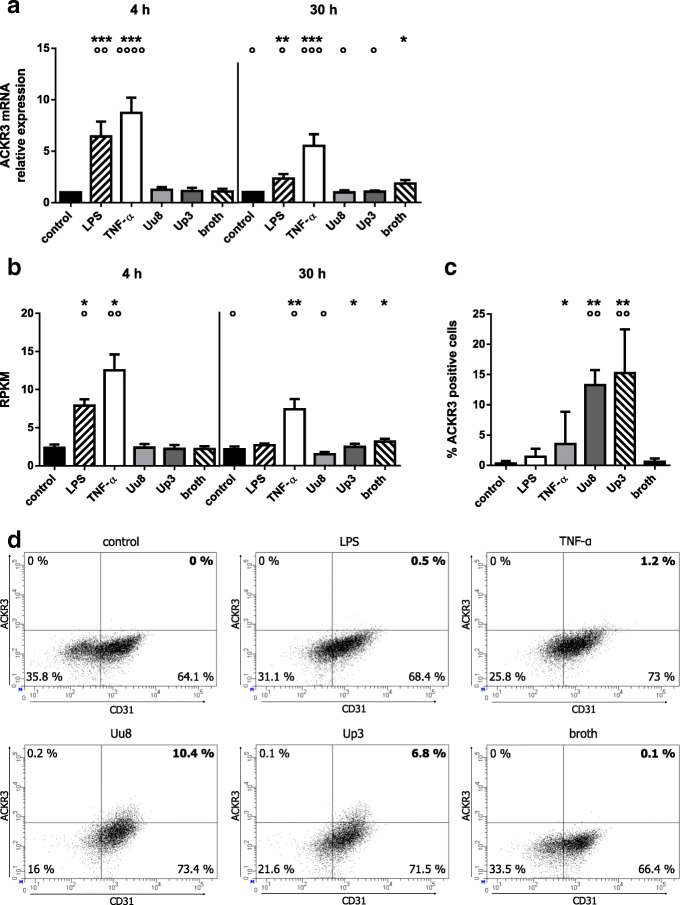
We established a cell culture model of Ureaplasma meningitis, aiming to analyze ACKR3 variances as a possible pathomechanism in Ureaplasma-associated neuroinflammation. Non-immortalized human brain microvascular endothelial cells (HBMEC) were exposed to bacterial lipopolysaccharide (LPS) or tumor necrosis factor-α (TNF-α), and native as well as LPS-primed HBMEC were cultured with Ureaplasma urealyticum serovar 8 (Uu8) and U. parvum serovar 3 (Up3). ACKR3 responses were assessed via qRT-PCR, RNA sequencing, flow cytometry, and immunocytochemistry.
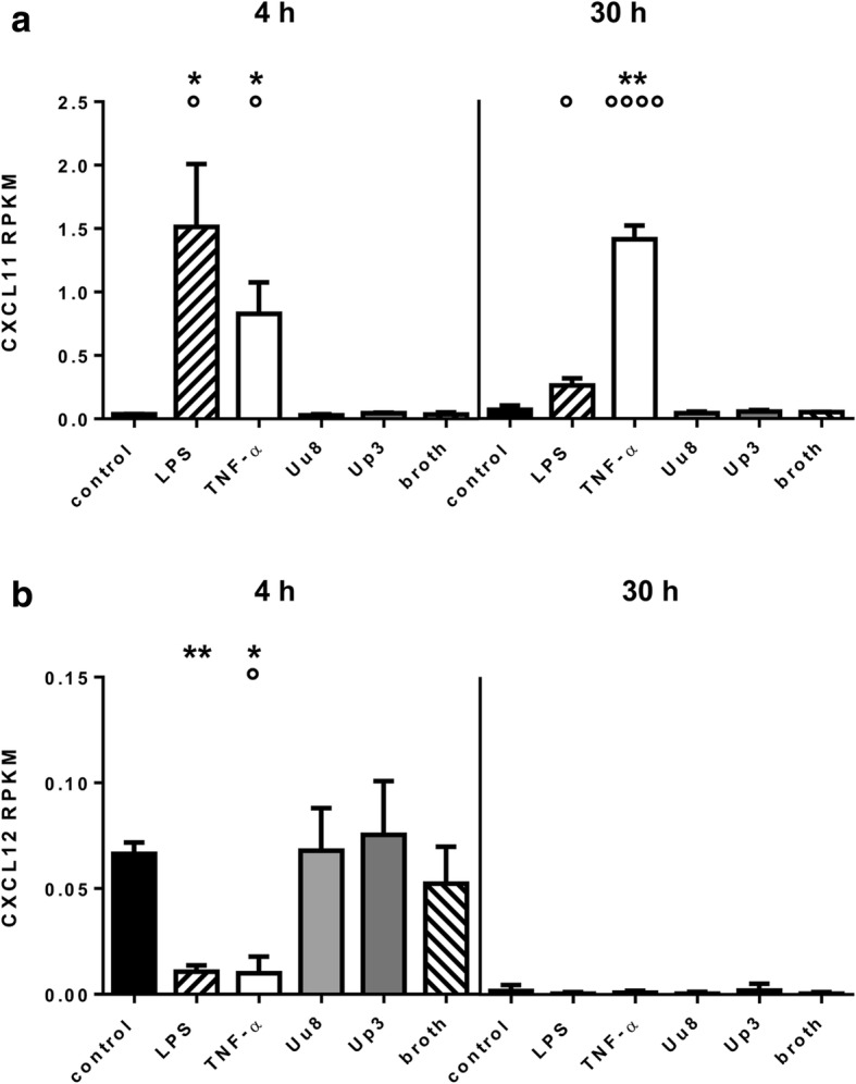
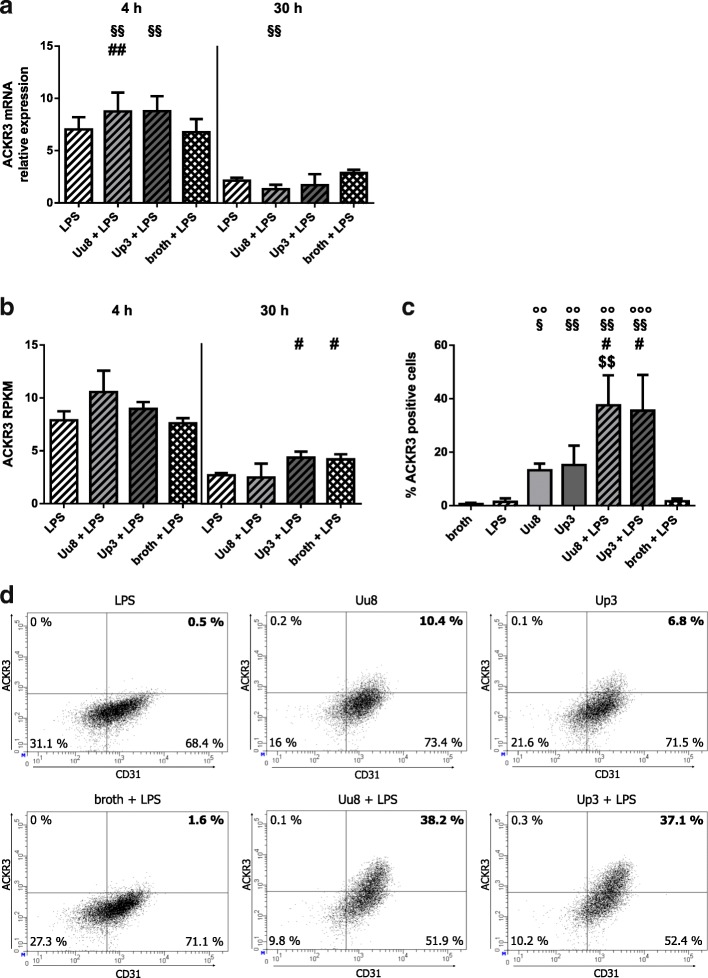
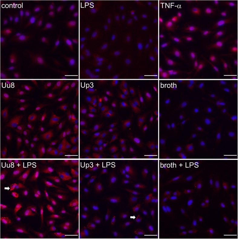
LPS, TNF-α, and Ureaplasma spp. influenced ACKR3 expression in HBMEC. LPS and TNF-α significantly induced ACKR3 mRNA expression (p < 0.001, vs. control), whereas Ureaplasma spp. enhanced ACKR3 protein expression in HBMEC (p < 0.01, vs. broth control). Co-stimulation with LPS and either Ureaplasma isolate intensified ACKR3 responses (p < 0.05, vs. LPS). Furthermore, stimulation wielded a differential influence on the receptor's ligands.
We introduce an in vitro model of Ureaplasma meningitis. We are able to demonstrate a pro-inflammatory capacity of Ureaplasma spp. in native and, even more so, in LPS-primed HBMEC, underlining their clinical relevance particularly in a setting of co-infection. Furthermore, our data may indicate a novel role for ACKR3, with an impact not limited to auto-inflammatory diseases, but extending to infection-related neuroinflammation as well. AKCR3-induced blood-brain barrier breakdown might constitute a potential common pathomechanism.
非典型趋化因子受体 3(ACKR3,同义词 CXCR7)在神经炎症性疾病中越来越受到重视,其上调导致内皮屏障功能受损,并可能最终导致炎症性脑损伤。虽然 ACKR3 的影响已在几种神经自身免疫性疾病中得到认可,但神经炎症也可能由包括解脲脲原体(Ureaplasma species,spp.)在内的感染因子引起。尽管解脲脲原体通常被认为是成人泌尿生殖道的共生菌,但它也可能在免疫功能低下的成人以及新生儿中引起侵袭性感染,并且似乎是新生儿脑膜炎的相关病原体。然而,关于解脲脲原体引起的炎症的临床和体外数据仍然很少。
我们建立了解脲脲原体脑膜炎的细胞培养模型,旨在分析 ACKR3 变异作为解脲脲原体相关神经炎症中的可能发病机制。非永生化人脑微血管内皮细胞(HBMEC)暴露于细菌脂多糖(LPS)或肿瘤坏死因子-α(TNF-α),并用解脲脲原体血清型 8(Uu8)和 U. parvum 血清型 3(Up3)培养天然和 LPS 预刺激的 HBMEC。通过 qRT-PCR、RNA 测序、流式细胞术和免疫细胞化学评估 ACKR3 反应。
LPS、TNF-α 和解脲脲原体 spp. 影响 HBMEC 中的 ACKR3 表达。LPS 和 TNF-α 显著诱导 ACKR3 mRNA 表达(p<0.001,与对照相比),而解脲脲原体 spp. 增强 HBMEC 中的 ACKR3 蛋白表达(p<0.01,与培养基对照相比)。LPS 和两种解脲脲原体分离株的共刺激增强了 ACKR3 反应(p<0.05,与 LPS 相比)。此外,刺激对受体配体产生了不同的影响。
我们引入了一种解脲脲原体脑膜炎的体外模型。我们能够证明解脲脲原体 spp. 在天然和 LPS 预刺激的 HBMEC 中具有促炎能力,突出了它们在合并感染中的临床相关性,特别是在合并感染的情况下。此外,我们的数据可能表明 ACKR3 具有新的作用,其影响不仅限于自身炎症性疾病,还扩展到与感染相关的神经炎症。ACKR3 诱导的血脑屏障破坏可能构成潜在的共同发病机制。