University Children's Hospital, University of Wuerzburg, Josef-Schneider-Str. 2, 97080, Wuerzburg, Germany.
Department of Pediatrics, Faculty of Health, Medicine and Life Sciences, Maastricht University Medical Center, P. Debyelaan 25, 6229 HX, Maastricht, Netherlands.
Cell Mol Neurobiol. 2023 Mar;43(2):785-795. doi: 10.1007/s10571-022-01213-8. Epub 2022 Mar 25.
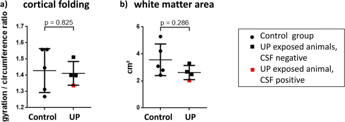
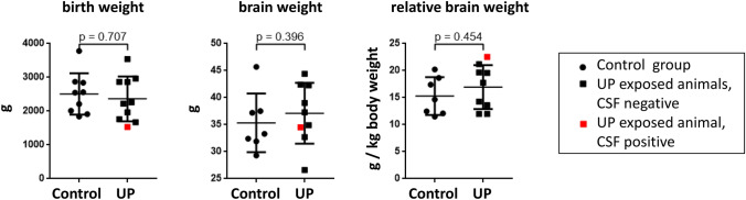
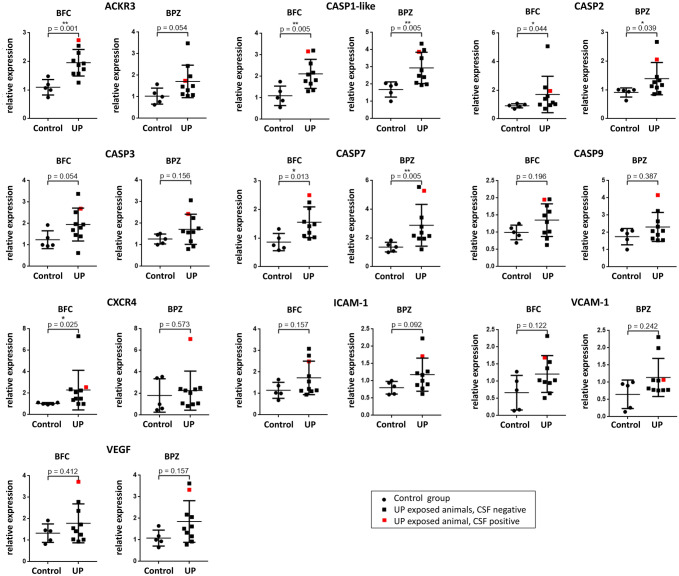
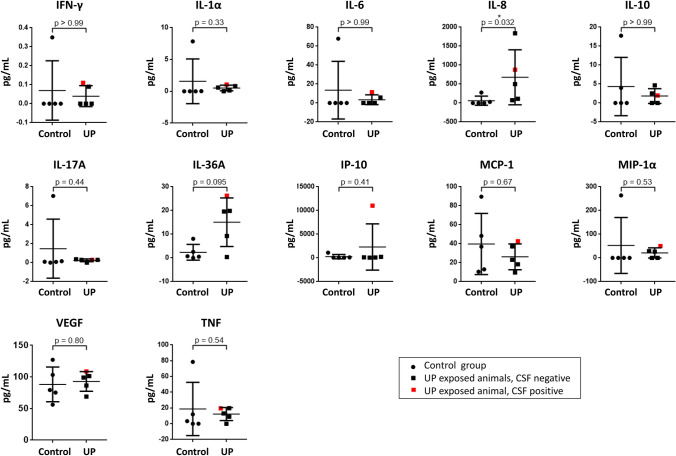
Ureaplasma species (spp.) are considered commensals of the adult genitourinary tract, but have been associated with chorioamnionitis, preterm birth, and invasive infections in neonates, including meningitis. Data on mechanisms involved in Ureaplasma-driven neuroinflammation are scarce. The present study addressed brain inflammatory responses in preterm lambs exposed to Ureaplasma parvum (UP) in utero. 7 days after intra-amniotic injection of UP (n = 10) or saline (n = 11), lambs were surgically delivered at gestational day 128-129. Expression of inflammatory markers was assessed in different brain regions using qRT-PCR and in cerebrospinal fluid (CSF) by multiplex immunoassay. CSF was analyzed for UP presence using ureB-based real-time PCR, and MRI scans documented cerebral white matter area and cortical folding. Cerebral tissue levels of atypical chemokine receptor (ACKR) 3, caspases 1-like, 2, 7, and C-X-C chemokine receptor (CXCR) 4 mRNA, as well as CSF interleukin-8 protein concentrations were significantly increased in UP-exposed lambs. UP presence in CSF was confirmed in one animal. Cortical folding and white matter area did not differ among groups. The present study confirms a role of caspases and the transmembrane receptors ACKR3 and CXCR4 in Ureaplasma-driven neuroinflammation. Enhanced caspase 1-like, 2, and 7 expression may reflect cell death. Increased ACKR3 and CXCR4 expression has been associated with inflammatory central nervous system (CNS) diseases and impaired blood-brain barrier function. According to these data and previous in vitro findings from our group, we speculate that Ureaplasma-induced caspase and receptor responses affect CNS barrier properties and thus facilitate neuroinflammation.
脲原体(Ureaplasma)种属被认为是成人泌尿生殖道的共生体,但与绒毛膜羊膜炎、早产和新生儿侵袭性感染有关,包括脑膜炎。关于脲原体驱动神经炎症的机制数据很少。本研究旨在探讨宫内暴露于微小脲原体(UP)的早产羔羊的脑炎症反应。在宫内注射 UP(n=10)或生理盐水(n=11)7 天后,于妊娠第 128-129 天进行手术分娩。通过 qRT-PCR 在不同脑区评估炎症标志物的表达,并通过多重免疫测定法在脑脊液(CSF)中评估。使用 ureB 基实时 PCR 检测 CSF 中 UP 的存在,MRI 扫描记录脑白质面积和皮质折叠。与对照组相比,暴露于 UP 的羔羊脑组织中趋化因子受体(ACKR)3、半胱氨酸蛋白酶 1 样、2、7 和 C-X-C 趋化因子受体(CXCR)4 mRNA 以及 CSF 白细胞介素-8 蛋白浓度显著增加。在一只动物的 CSF 中证实了 UP 的存在。各组皮质折叠和白质面积无差异。本研究证实了半胱氨酸蛋白酶和跨膜受体 ACKR3 和 CXCR4 在 UP 驱动的神经炎症中的作用。增强的半胱氨酸蛋白酶 1 样、2 和 7 表达可能反映细胞死亡。ACKR3 和 CXCR4 表达增加与炎症性中枢神经系统(CNS)疾病和血脑屏障功能受损有关。根据这些数据和我们小组以前的体外发现,我们推测 UP 诱导的半胱氨酸蛋白酶和受体反应影响 CNS 屏障特性,从而促进神经炎症。