Institute of Neuroscience, Medical School, Newcastle University, Newcastle upon Tyne NE2 4HH, UK.
Institute of Neuroscience, Medical School, Newcastle University, Newcastle upon Tyne NE2 4HH, UK
J Neurosci. 2018 Jul 4;38(27):6190-6206. doi: 10.1523/JNEUROSCI.3371-17.2018. Epub 2018 May 23.
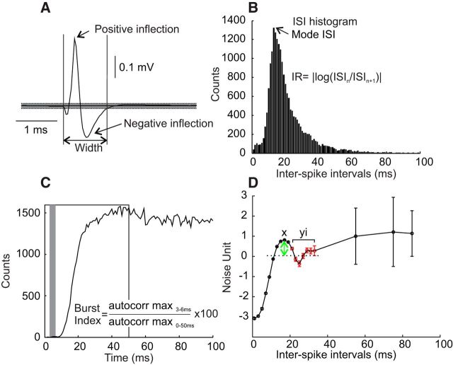
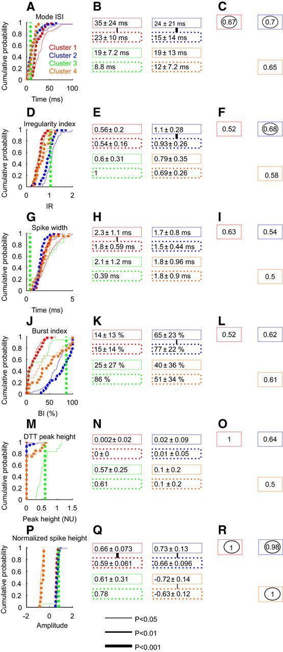
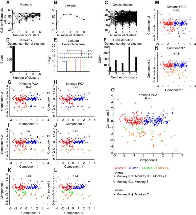
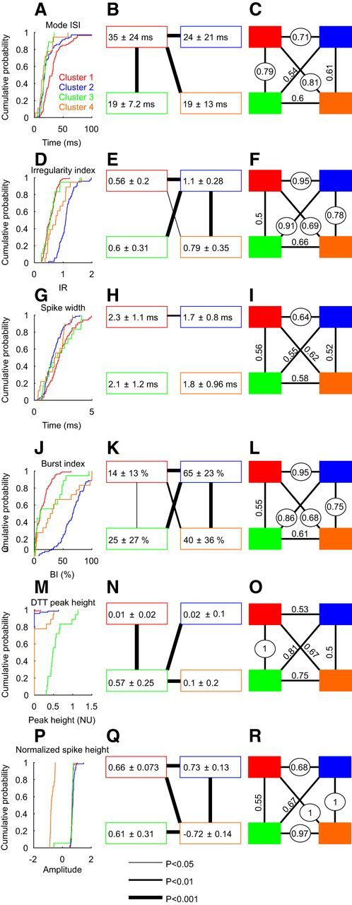
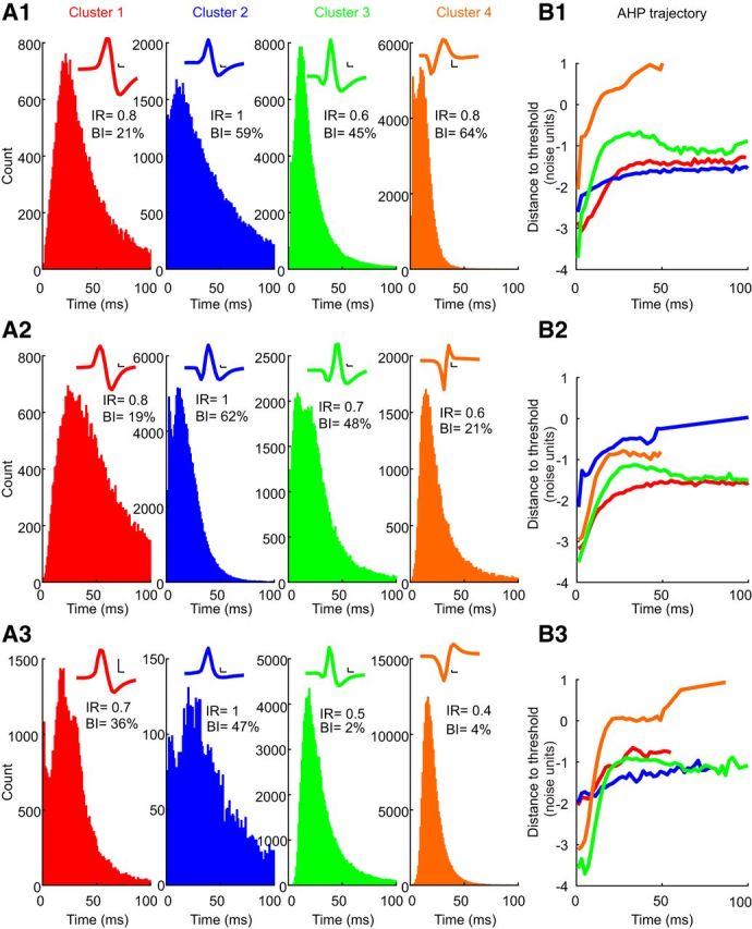
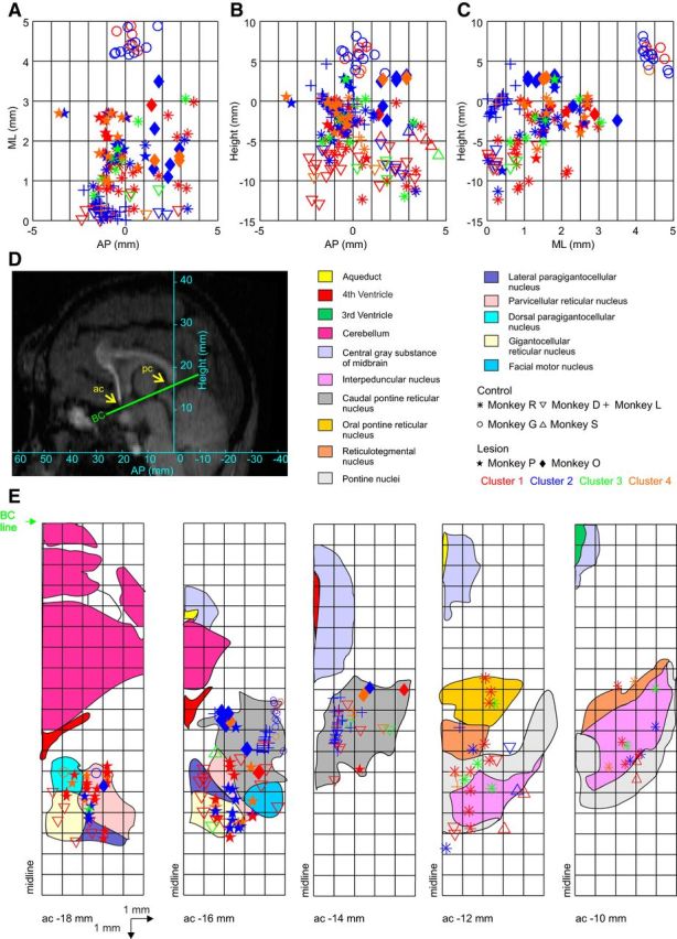
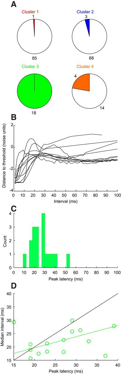
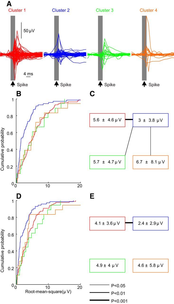
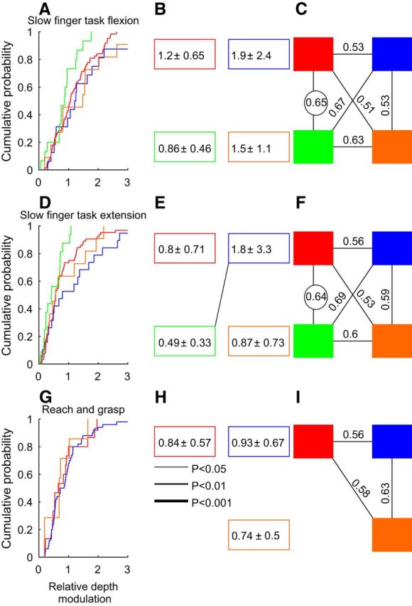
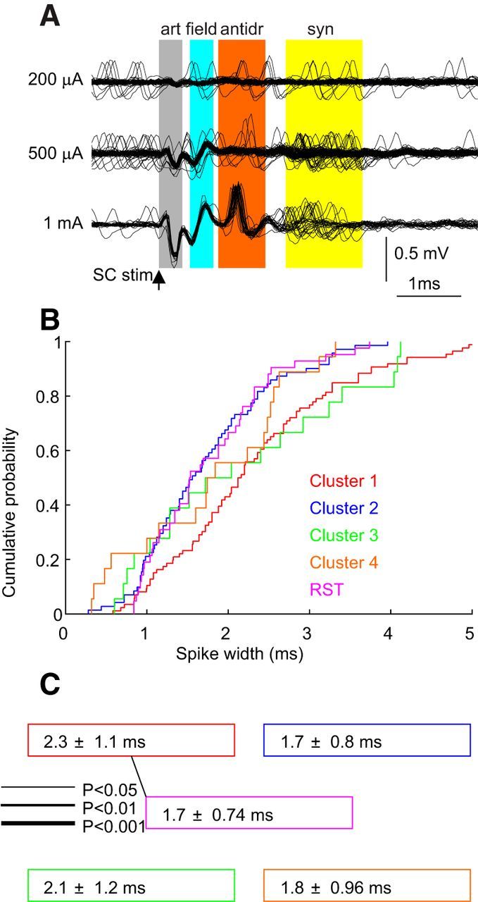
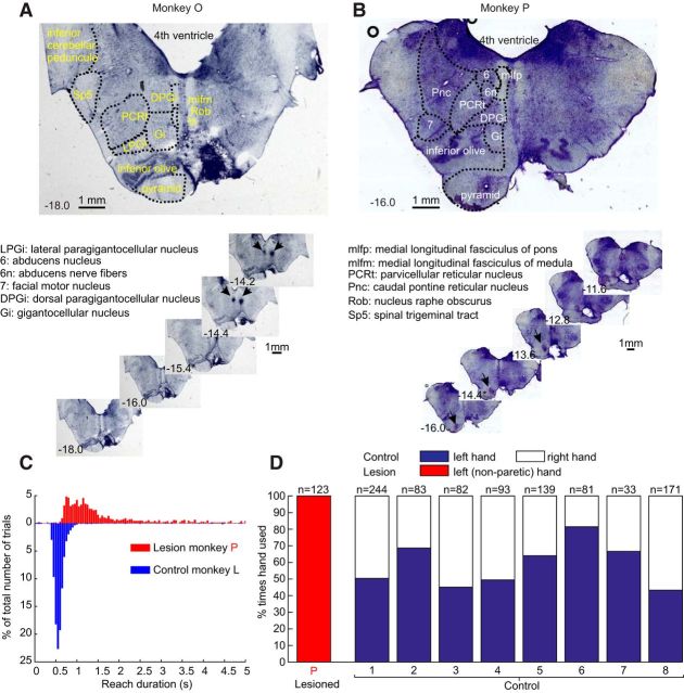
The reticular formation is important in primate motor control, both in health and during recovery after brain damage. Little is known about the different neurons present in the reticular nuclei. Here we recorded extracellular spikes from the reticular formation in five healthy female awake behaving monkeys (193 cells), and in two female monkeys 1 year after recovery from a unilateral pyramidal tract lesion (125 cells). Analysis of spike shape and four measures derived from the interspike interval distribution identified four clusters of neurons in control animals. Cluster 1 cells had a slow firing rate. Cluster 2 cells had narrow spikes and irregular firing, which often included high-frequency bursts. Cluster 3 cells were highly rhythmic and fast firing. Cluster 4 cells showed negative spikes. A separate population of 42 cells was antidromically identified as reticulospinal neurons in five anesthetized female monkeys. The distribution of spike width in these cells closely overlaid the distribution for cluster 2, leading us tentatively to suggest that cluster 2 included neurons with reticulospinal projections. In animals after corticospinal lesion, cells could be identified in all four clusters. The firing rate of cells in clusters 1 and 2 was increased in lesioned animals relative to control animals (by 52% and 60%, respectively); cells in cluster 2 were also more regular and more bursting in the lesioned animals. We suggest that changes in both membrane properties and local circuits within the reticular formation occur following lesioning, potentially increasing reticulospinal output to help compensate for lost corticospinal descending drive. This work is the first to subclassify neurons in the reticular formation, providing insights into the local circuitry of this important but little understood structure. The approach developed can be applied to any extracellular recording from this region, allowing future studies to place their data within our current framework of four neural types. Changes in reticular neurons may be important to subserve functional recovery after damage in human patients, such as after stroke or spinal cord injury.
网状结构在灵长类动物的运动控制中很重要,无论是在健康状态下还是在脑损伤后的恢复过程中。目前对于网状核内存在的不同神经元知之甚少。在这里,我们在五只健康的、清醒的、行为正常的雌性猴子(193 个细胞)中记录了网状结构的细胞外放电,还在两只雌性猴子从单侧锥体束损伤中恢复 1 年后(125 个细胞)记录了它们的细胞外放电。对尖峰形状和四个源自尖峰间隔分布的测量指标进行分析,在对照动物中确定了四个神经元簇。簇 1 细胞的放电频率较慢。簇 2 细胞的尖峰较窄,放电不规则,通常包括高频爆发。簇 3 细胞节律性强,放电速度快。簇 4 细胞表现出负尖峰。在五只麻醉雌性猴子中,通过对 42 个细胞进行逆行性识别,确定了一个单独的细胞群体为网状脊髓神经元。这些细胞的尖峰宽度分布与簇 2 的分布紧密重叠,这使我们推测簇 2 包括具有网状脊髓投射的神经元。在皮质脊髓束损伤后的动物中,可以在所有四个簇中识别出细胞。与对照动物相比,损伤动物中簇 1 和簇 2 细胞的放电频率增加(分别增加了 52%和 60%);损伤动物中簇 2 细胞的放电也更加规则,爆发性更强。我们认为,损伤后网状结构内的膜特性和局部回路都发生了变化,这可能增加了网状脊髓输出,有助于补偿失去的皮质脊髓下行驱动。这项工作首次对网状结构中的神经元进行了亚分类,为了解这一重要但了解甚少的结构的局部回路提供了深入的认识。所开发的方法可以应用于该区域的任何细胞外记录,从而使未来的研究能够将其数据置于我们当前的四种神经类型框架内。网状神经元的变化对于人类患者(如中风或脊髓损伤后)的功能恢复可能很重要。