• 文献检索
  • 文档翻译
  • 深度研究
  • 学术资讯
  • Suppr Zotero 插件Zotero 插件
  • 邀请有礼
  • 套餐&价格
  • 历史记录
应用&插件
Suppr Zotero 插件Zotero 插件浏览器插件Mac 客户端Windows 客户端微信小程序
定价
高级版会员购买积分包购买API积分包
服务
文献检索文档翻译深度研究API 文档MCP 服务
关于我们
关于 Suppr公司介绍联系我们用户协议隐私条款
关注我们

Suppr 超能文献

核心技术专利:CN118964589B侵权必究
粤ICP备2023148730 号-1Suppr @ 2026

文献检索

告别复杂PubMed语法,用中文像聊天一样搜索,搜遍4000万医学文献。AI智能推荐,让科研检索更轻松。

立即免费搜索

文件翻译

保留排版,准确专业,支持PDF/Word/PPT等文件格式,支持 12+语言互译。

免费翻译文档

深度研究

AI帮你快速写综述,25分钟生成高质量综述,智能提取关键信息,辅助科研写作。

立即免费体验

评估视网膜色素变性患者的中心视觉功能。

Assessment of Central Visual Function in Patients with Retinitis Pigmentosa.

机构信息

Department of Ophthalmology, Graduate School of Medical Sciences, Kyushu University, Fukuoka, 812-8582, Japan.

Department of Ophthalmology, Graduate School of Medical Sciences, Akita University, Akita, 010-8543, Japan.

出版信息

Sci Rep. 2018 May 23;8(1):8070. doi: 10.1038/s41598-018-26231-9.

DOI:10.1038/s41598-018-26231-9
PMID:29795288
原文链接:https://pmc.ncbi.nlm.nih.gov/articles/PMC5966451/
Abstract

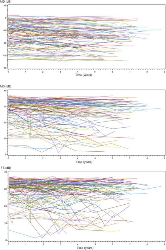
In order to clarify the disease progression in retinitis pigmentosa (RP) and its related factors, reliable data on the changes in central visual function in RP are needed. In this longitudinal study, we examined 118 patients who were diagnosed with typical RP. Visual acuity (VA), visual field using a Humphrey Field Analyzer with the central 10-2 SITA-Standard program, and optical coherence tomography measurements were obtained. The slopes, which were derived from serial values of mean deviation (MD), macular sensitivity (MS), or foveal sensitivity (FS) obtained for each eye by a linear mixed model, were used for analysis. MS and FS were calculated as the average retinal sensitivity of 12 and 4 central points respectively. There were statistically significant interactions of times with levels of the central subfield thickness (CST) on the slopes of MS and FS. Compared to the eyes without macular complications, the eyes with macular complications had steeper MD, MS and FS slopes, and this interaction was no significant, but marginal trend for the MS or FS slope (P = 0.10, 0.05, respectively). The central retinal sensitivity (i.e., MS and FS) slopes calculated were effective indices of the progression of central visual function in RP.

摘要

为了阐明色素性视网膜炎(RP)的疾病进展及其相关因素,需要可靠的数据来反映 RP 患者中心视力功能的变化。在这项纵向研究中,我们检查了 118 名被诊断为典型 RP 的患者。使用 Humphrey 视野分析仪(中央 10-2 SITA-Standard 程序)获取了视力(VA)、视野和光相干断层扫描(OCT)测量值。通过线性混合模型获得每个眼睛的平均偏差(MD)、黄斑敏感性(MS)或中央敏感性(FS)的系列值,使用斜率来进行分析。MS 和 FS 分别计算为 12 个和 4 个中央点的视网膜平均敏感性。MS 和 FS 斜率与中央子场厚度(CST)水平之间存在统计学显著的时间交互作用。与没有黄斑并发症的眼睛相比,有黄斑并发症的眼睛的 MD、MS 和 FS 斜率更陡峭,尽管这种交互作用对于 MS 或 FS 斜率没有显著意义,但呈边际趋势(P=0.10,0.05)。计算得出的中央视网膜敏感性(即 MS 和 FS)斜率是 RP 中心视力功能进展的有效指标。

https://cdn.ncbi.nlm.nih.gov/pmc/blobs/5668/5966451/d76a38f2d192/41598_2018_26231_Fig2_HTML.jpg
https://cdn.ncbi.nlm.nih.gov/pmc/blobs/5668/5966451/54802c5a5656/41598_2018_26231_Fig1_HTML.jpg
https://cdn.ncbi.nlm.nih.gov/pmc/blobs/5668/5966451/d76a38f2d192/41598_2018_26231_Fig2_HTML.jpg
https://cdn.ncbi.nlm.nih.gov/pmc/blobs/5668/5966451/54802c5a5656/41598_2018_26231_Fig1_HTML.jpg
https://cdn.ncbi.nlm.nih.gov/pmc/blobs/5668/5966451/d76a38f2d192/41598_2018_26231_Fig2_HTML.jpg

相似文献

1
Assessment of Central Visual Function in Patients with Retinitis Pigmentosa.评估视网膜色素变性患者的中心视觉功能。
Sci Rep. 2018 May 23;8(1):8070. doi: 10.1038/s41598-018-26231-9.
2
FEATURES OF THE MACULA AND CENTRAL VISUAL FIELD AND FIXATION PATTERN IN PATIENTS WITH RETINITIS PIGMENTOSA.色素性视网膜炎患者的黄斑和中央视野及固视模式特征。
Retina. 2018 Feb;38(2):424-431. doi: 10.1097/IAE.0000000000001532.
3
Optical coherence tomography angiography of the macular microvasculature changes in retinitis pigmentosa.视网膜色素变性中黄斑部微血管变化的光学相干断层扫描血管造影
Acta Ophthalmol. 2018 Feb;96(1):e59-e67. doi: 10.1111/aos.13475. Epub 2017 May 31.
4
Aqueous Flare and Progression of Visual Field Loss in Patients With Retinitis Pigmentosa.色素性视网膜炎患者的房水闪光和视野丧失进展。
Invest Ophthalmol Vis Sci. 2020 Jul 1;61(8):26. doi: 10.1167/iovs.61.8.26.
5
The qualitative assessment of optical coherence tomography and the central retinal sensitivity in patients with retinitis pigmentosa.视网膜色素变性患者的光学相干断层扫描和中心视网膜敏感度的定性评估。
PLoS One. 2020 May 11;15(5):e0232700. doi: 10.1371/journal.pone.0232700. eCollection 2020.
6
Correlation between macular blood flow and central visual sensitivity in retinitis pigmentosa.视网膜色素变性患者黄斑血流与中心视觉敏感度之间的相关性
Acta Ophthalmol. 2015 Dec;93(8):e644-8. doi: 10.1111/aos.12693. Epub 2015 Feb 17.
7
Correlation between aqueous flare and residual visual field area in retinitis pigmentosa.色素性视网膜炎中水闪辉与残余视野面积的相关性。
Br J Ophthalmol. 2019 Apr;103(4):475-480. doi: 10.1136/bjophthalmol-2018-312225. Epub 2018 Jun 1.
8
Correlation between Visual Function and Structural Characteristics of the Macula in Advanced Retinitis Pigmentosa.晚期视网膜色素变性的视功能与黄斑区结构特征的相关性。
Ophthalmologica. 2019;242(1):22-30. doi: 10.1159/000496941. Epub 2019 Mar 20.
9
Relations Among Foveal Blood Flow, Retinal-Choroidal Structure, and Visual Function in Retinitis Pigmentosa.色素性视网膜炎中黄斑血流、视网膜脉络膜结构与视觉功能的关系。
Invest Ophthalmol Vis Sci. 2018 Feb 1;59(2):1134-1143. doi: 10.1167/iovs.17-23050.
10
The clinical efficacy of a topical dorzolamide in the management of cystoid macular edema in patients with retinitis pigmentosa.局部多佐胺治疗色素性视网膜炎患者囊样黄斑水肿的临床疗效。
Graefes Arch Clin Exp Ophthalmol. 2012 Jun;250(6):809-14. doi: 10.1007/s00417-011-1904-5. Epub 2012 Jan 4.

引用本文的文献

1
Study protocol for a prospective natural history registry investigating the relationships between inflammatory markers and disease progression in retinitis pigmentosa: the RP-PRIMARY study.一项前瞻性自然史登记研究的研究方案,该研究旨在调查色素性视网膜炎中炎症标志物与疾病进展之间的关系:RP-PRIMARY研究。
Jpn J Ophthalmol. 2025 Mar 5. doi: 10.1007/s10384-025-01179-2.
2
Good vision without peripheries: behavioral and fMRI evidence.良好的中心视力而周边视力不佳:行为和 fMRI 的证据。
Sci Rep. 2024 Nov 1;14(1):26264. doi: 10.1038/s41598-024-76879-9.
3
Comparison of Microperimetry and Static Perimetry for Evaluating Macular Function and Progression in Retinitis Pigmentosa.

本文引用的文献

1
Longitudinal study of visual field changes determined by Humphrey Field Analyzer 10-2 in patients with Retinitis Pigmentosa.视网膜色素变性患者 Humphrey 视野分析仪 10-2 检测的视野变化的纵向研究。
Sci Rep. 2017 Nov 27;7(1):16383. doi: 10.1038/s41598-017-16640-7.
2
Mutation screening in genes known to be responsible for Retinitis Pigmentosa in 98 Small Han Chinese Families.在中国 98 个小汉族家庭中,对已知与色素性视网膜炎相关的基因进行突变筛查。
Sci Rep. 2017 May 16;7(1):1948. doi: 10.1038/s41598-017-00963-6.
3
Risk Factors for Posterior Subcapsular Cataract in Retinitis Pigmentosa.
微视野检查法与静态视野检查法在评估视网膜色素变性黄斑功能及病情进展中的比较
Ophthalmol Sci. 2024 Jul 20;4(6):100582. doi: 10.1016/j.xops.2024.100582. eCollection 2024 Nov-Dec.
4
Progression Rate of Visual Function and Affecting Factors at Different Stages of Retinitis Pigmentosa.色素性视网膜炎不同阶段的视觉功能进展率及其影响因素。
Biomed Res Int. 2022 Jul 14;2022:7204954. doi: 10.1155/2022/7204954. eCollection 2022.
5
Real-world outcomes of voretigene neparvovec treatment in pediatric patients with RPE65-associated Leber congenital amaurosis.经 RPE65 相关莱伯先天性黑矇治疗的儿科患者中 voretigene neparvovec 治疗的真实世界结局。
Graefes Arch Clin Exp Ophthalmol. 2022 May;260(5):1543-1550. doi: 10.1007/s00417-021-05508-2. Epub 2022 Jan 10.
6
Development and validation of a visual field cluster in retinitis pigmentosa.开发和验证视网膜色素变性的视野簇。
Sci Rep. 2021 May 6;11(1):9671. doi: 10.1038/s41598-021-89233-0.
7
Changes of Serum Inflammatory Molecules and Their Relationships with Visual Function in Retinitis Pigmentosa.色素性视网膜炎患者血清炎症分子的变化及其与视觉功能的关系。
Invest Ophthalmol Vis Sci. 2020 Sep 1;61(11):30. doi: 10.1167/iovs.61.11.30.
8
Aqueous Flare and Progression of Visual Field Loss in Patients With Retinitis Pigmentosa.色素性视网膜炎患者的房水闪光和视野丧失进展。
Invest Ophthalmol Vis Sci. 2020 Jul 1;61(8):26. doi: 10.1167/iovs.61.8.26.
9
The qualitative assessment of optical coherence tomography and the central retinal sensitivity in patients with retinitis pigmentosa.视网膜色素变性患者的光学相干断层扫描和中心视网膜敏感度的定性评估。
PLoS One. 2020 May 11;15(5):e0232700. doi: 10.1371/journal.pone.0232700. eCollection 2020.
10
Metabolic and Redox Signaling of the Nucleoredoxin-Like-1 Gene for the Treatment of Genetic Retinal Diseases.核蛋白样 1 基因的代谢和氧化还原信号转导在遗传性视网膜疾病治疗中的作用。
Int J Mol Sci. 2020 Feb 27;21(5):1625. doi: 10.3390/ijms21051625.
视网膜色素变性患者后囊下白内障的危险因素
Invest Ophthalmol Vis Sci. 2017 May 1;58(5):2534-2537. doi: 10.1167/iovs.17-21612.
4
Association Between Aqueous Flare and Epiretinal Membrane in Retinitis Pigmentosa.色素性视网膜炎中房水闪光与视网膜前膜之间的关联
Invest Ophthalmol Vis Sci. 2016 Aug 1;57(10):4282-6. doi: 10.1167/iovs.16-19686.
5
Neuoroprotective efficacies by KUS121, a VCP modulator, on animal models of retinal degeneration.VCP调节剂KUS121对视网膜变性动物模型的神经保护作用。
Sci Rep. 2016 Aug 9;6:31184. doi: 10.1038/srep31184.
6
Correlation of structure and function of the macula in patients with retinitis pigmentosa.视网膜色素变性患者黄斑区结构与功能的相关性
Eye (Lond). 2015 Jul;29(7):895-901. doi: 10.1038/eye.2015.61. Epub 2015 May 8.
7
Relationship between aqueous flare and visual function in retinitis pigmentosa.色素性视网膜炎中房水闪光与视觉功能的关系。
Am J Ophthalmol. 2015 May;159(5):958-63.e1. doi: 10.1016/j.ajo.2015.02.001. Epub 2015 Feb 10.
8
Therapeutic efficacy of topical unoprostone isopropyl in retinitis pigmentosa.局部用异丙前列素治疗色素性视网膜炎的疗效
Acta Ophthalmol. 2014 May;92(3):e229-34. doi: 10.1111/aos.12293.
9
Progress of mesenchymal stem cell therapy for neural and retinal diseases.间充质干细胞治疗神经和视网膜疾病的研究进展
World J Stem Cells. 2014 Apr 26;6(2):111-9. doi: 10.4252/wjsc.v6.i2.111.
10
Macular abnormalities in Italian patients with retinitis pigmentosa.意大利色素性视网膜炎患者的黄斑异常。
Br J Ophthalmol. 2014 Jul;98(7):946-50. doi: 10.1136/bjophthalmol-2013-304082. Epub 2014 Feb 13.