Athinoula A. Martinos Center for Biomedical Imaging, Massachusetts General Hospital and Harvard Medical School, Boston, MA, 02129, USA.
School of Pharmacy, China Pharmaceutical University, Nanjing, 210009, China.
Mol Imaging Biol. 2019 Feb;21(1):35-43. doi: 10.1007/s11307-018-1213-z.
Near-infrared fluorescence (NIRF) imaging has been widely used in preclinical studies; however, its low tissue penetration represents a daunting problem for translational clinical imaging of neurodegenerative diseases. The retina is known as an extension of the central nerve system (CNS), and it is widely considered as a window to the brain. Therefore, the retina can be considered as an alternative organ for investigating neurodegenerative diseases, and an eye represents an ideal NIRF imaging organ, due to its minimal opacity.
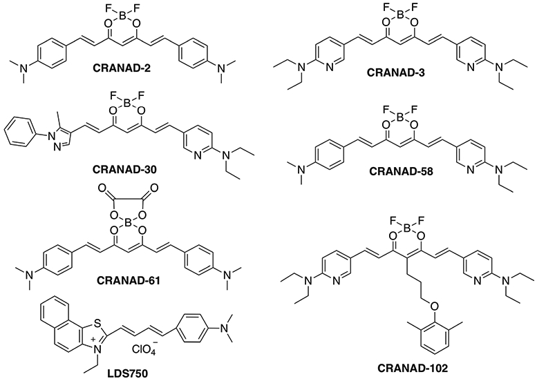
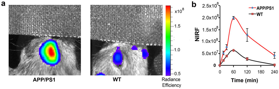
NIRF ocular imaging (NIRFOI), for the first time, was explored for imaging of Alzheimer's disease (AD) via utilizing "smart" fluorescent probes CRANAD-X (X = - 2, - 3, - 30, - 58, and - 102) for amyloid beta (Aβ), and CRANAD-61 for reactive oxygen species (ROS). Mice were intravenously injected the fluorescence dyes and images from the eyes were captured with an IVIS imaging system at different time points.
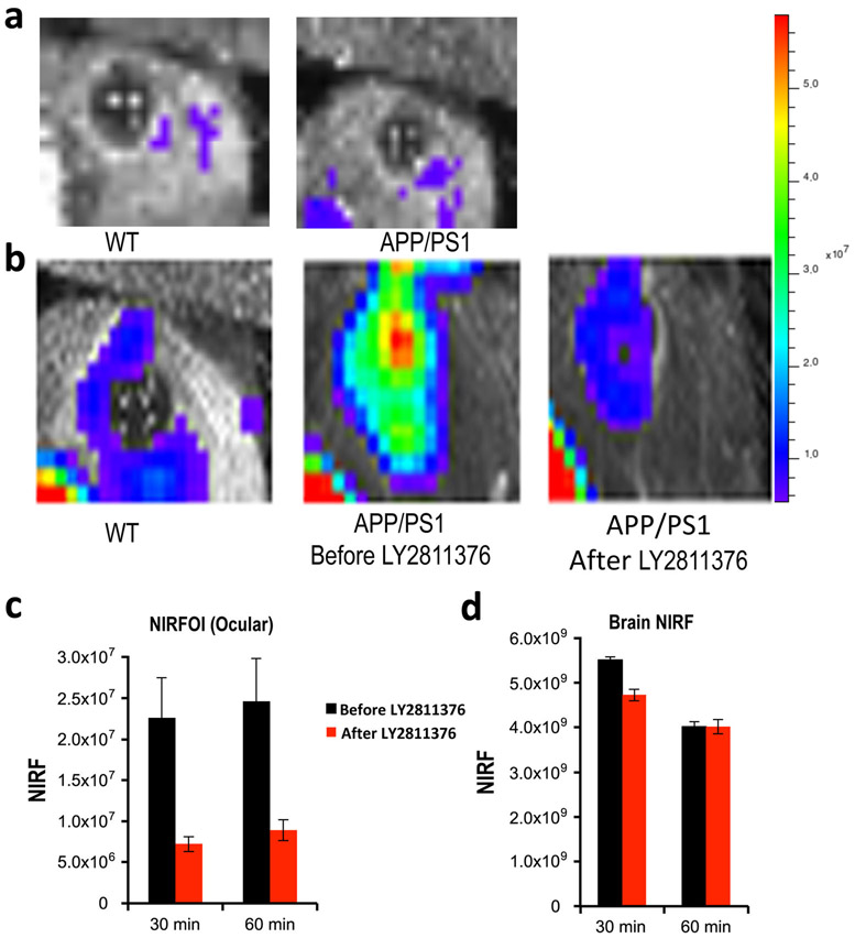
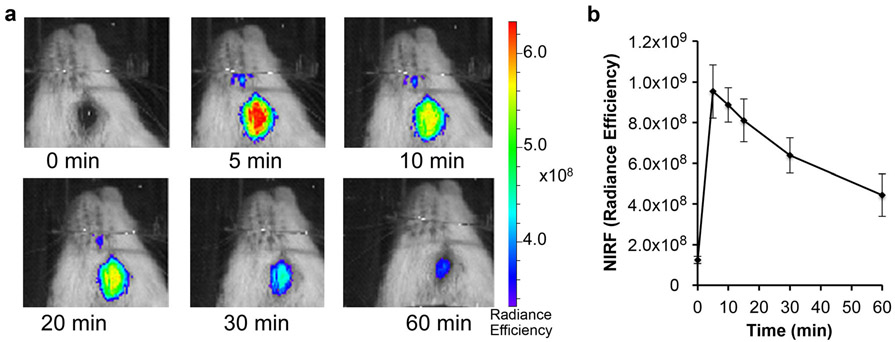
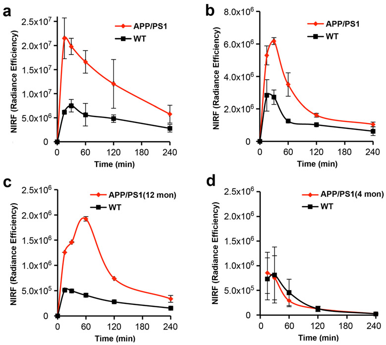
All of the tested NIRF probes could be used to differentiate transgenic AD mice and WT mice, and NIRFOI could provide much higher sensitivity for imaging Aβs than NIRF brain imaging did. Our data suggested that NIRFOI could capture the imaging signals from both soluble and insoluble Aβ species. Moreover, we demonstrated that NIRFOI with CRANAD-102 could be used to monitor the therapeutic effects of BACE-1 inhibitor LY2811376. Compared to NIRF brain imaging, NIRFOI provided a larger change of Aβ levels before and after LY2811376 treatment. In addition, we demonstrated that CRANAD-61 could be used to image reactive oxygen species in the eyes.
The large detection margin by NIRFOI is very important for both diagnosis and therapy response monitoring. Compared to fluorescence microscopic imaging, NIRFOI captures signals with a wide angle (large field of view (FOV)) and can be used to detect soluble Aβs. We believe that NIRFOI has remarkable translational potential for future human studies and can be a potential imaging technology for fast, cheap, accessible, and reliable screening of AD in the future.
近红外荧光(NIRF)成像已广泛应用于临床前研究;然而,其组织穿透深度低,这是神经退行性疾病转化临床成像的一个令人畏惧的问题。视网膜被认为是中枢神经系统(CNS)的延伸,它通常被认为是大脑的窗口。因此,视网膜可以被视为研究神经退行性疾病的替代器官,由于其最小的不透明度,眼睛是进行 NIRF 成像的理想器官。
首次通过利用“智能”荧光探针 CRANAD-X(X=−2、−3、−30、−58 和−102)检测淀粉样β(Aβ)和 CRANAD-61 检测活性氧(ROS),对 NIRF 眼部成像(NIRFOI)进行了探索,以进行阿尔茨海默病(AD)成像。将荧光染料静脉内注射到小鼠体内,并在不同时间点使用 IVIS 成像系统捕获眼睛的图像。
所有测试的 NIRF 探针都可用于区分转 AD 基因的小鼠和 WT 小鼠,与 NIRF 脑成像相比,NIRFOI 对 Aβ的成像灵敏度更高。我们的数据表明,NIRFOI 可以捕获可溶性和不溶性 Aβ 物种的成像信号。此外,我们证明了 NIRFOI 与 CRANAD-102 结合可用于监测 BACE-1 抑制剂 LY2811376 的治疗效果。与 NIRF 脑成像相比,LY2811376 治疗前后 NIRFOI 提供的 Aβ 水平变化更大。此外,我们证明了 CRANAD-61 可用于眼部活性氧成像。
NIRFOI 的大检测范围对于诊断和治疗反应监测都非常重要。与荧光显微镜成像相比,NIRFOI 以宽视角(大视野(FOV))捕获信号,并且可用于检测可溶性 Aβ。我们相信,NIRFOI 具有显著的转化潜力,可用于未来的人类研究,并且可能成为未来 AD 快速、廉价、可及且可靠筛查的潜在成像技术。