Ghaderi Hamid, Razmkhah Mahboobeh, Kiany Farin, Chenari Nooshafarin, Haghshenas Mohammad Reza, Ghaderi Abbas
Dentist, Private Practice, Shiraz, Iran.
Institute for Cancer Research, School of Medicine, Shiraz University of Medical Sciences, Shiraz, Iran.
J Dent (Shiraz). 2018 Jun;19(2):124-131.
One major goal of tissue engineering and regenerative medicine is to find an appropriate source of mesenchymal stem cells (MSCs) with higher differentiation ability.
In this experimental study, the osteogenic and chondrogenic differentiation ability of buccal fat pad derived MSCs (BFP-MSCs) with gingival derived cells (GDCs) were compared.
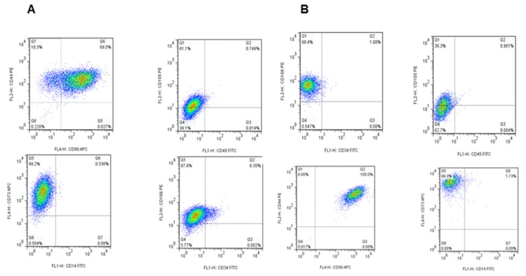
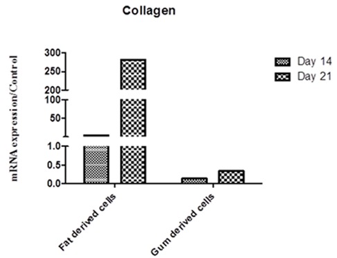
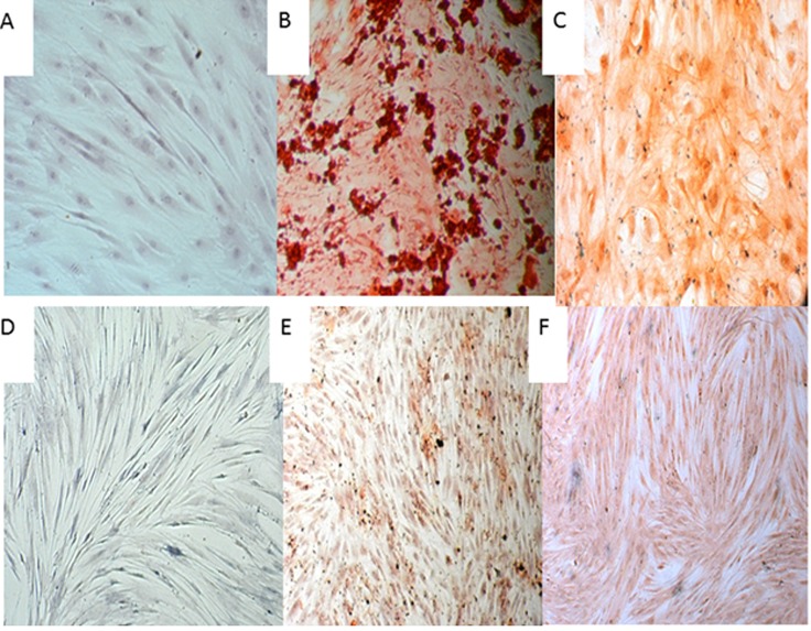
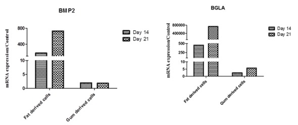
BFP-MSCs and GDCs were cultured enzymatically and expanded. The expanded cells were analyzed for membrane-associated markers, using flow cytometry. Then the ability of these cells to differentiate into osteocyte and chondrocyte was assessed morphologically and by mRNA expression of collagen I (COLL), BGLA and bone morphogenetic protein 2 (BMP2) using qRT-PCR.
Flow cytometry analysis showed that both BFP-MSCs and GDCs expressed the characteristic stem cell markers such as CD73, CD44, and CD90, whereas they did not express hematopoietic markers. Mineralized calcium deposition was observed apparently in BFP-MSCs cultured in osteogenic medium but GDCs showed fewer mineralized nodules. The mRNA expression levels of BGLA and BMP2 showed 7×10⁵ and 733-fold more mRNA expression in BFP-MSCs treated with differentiation media compared to the control group. In chondrogenic differentiation, BFP-MSCs transformed from a spindle to a cuboidal shape while GDCs showed only a slight transformation. In addition, mRNA expression of COLL showed 282-fold higher expression in BFP-MSCs in comparison to the control group. Such significant difference in mRNA expression of BGLA, BMP2, and COLL was not observed in GDCs compared to their corresponding controls.
Based on the present results, BFP yields a greater proportion of stem cells compared to gingiva. Therefore, this tissue can be introduced as an easily available source for the treatment of periodontal defects and other maxillofacial injuries.
组织工程和再生医学的一个主要目标是找到具有更高分化能力的间充质干细胞(MSC)的合适来源。
在本实验研究中,比较了颊脂垫来源的间充质干细胞(BFP-MSC)与牙龈来源细胞(GDC)的成骨和成软骨分化能力。
酶解培养并扩增BFP-MSC和GDC。使用流式细胞术分析扩增细胞的膜相关标志物。然后通过形态学评估这些细胞分化为骨细胞和软骨细胞的能力,并使用qRT-PCR通过胶原蛋白I(COLL)、BGLA和骨形态发生蛋白2(BMP2)的mRNA表达进行评估。
流式细胞术分析表明,BFP-MSC和GDC均表达特征性干细胞标志物,如CD73、CD44和CD90,而它们不表达造血标志物。在成骨培养基中培养的BFP-MSC中明显观察到矿化钙沉积,但GDC显示出较少的矿化结节。与对照组相比,用分化培养基处理的BFP-MSC中BGLA和BMP2的mRNA表达水平分别高7×10⁵倍和733倍。在软骨分化中,BFP-MSC从纺锤形转变为立方形,而GDC仅显示轻微转变。此外,与对照组相比,BFP-MSC中COLL的mRNA表达高282倍。与相应对照组相比,GDC中未观察到BGLA、BMP2和COLL的mRNA表达有如此显著差异。
基于目前的结果,与牙龈相比,颊脂垫产生的干细胞比例更高。因此,该组织可作为治疗牙周缺损和其他颌面损伤的容易获得的来源。