Dohmen Amy J C, Sanders Joyce, Canisius Sander, Jordanova Ekaterina S, Aalbersberg Else A, van den Brekel Michiel W M, Neefjes Jacques, Zuur Charlotte L
Department of Head and Neck Oncology and Surgery, Antoni van Leeuwenhoek, Amsterdam, The Netherlands.
Department of Pathology, the Netherlands Cancer Institute - Antoni van Leeuwenhoek, Amsterdam, The Netherlands.
Oncotarget. 2018 May 18;9(38):25034-25047. doi: 10.18632/oncotarget.25244.
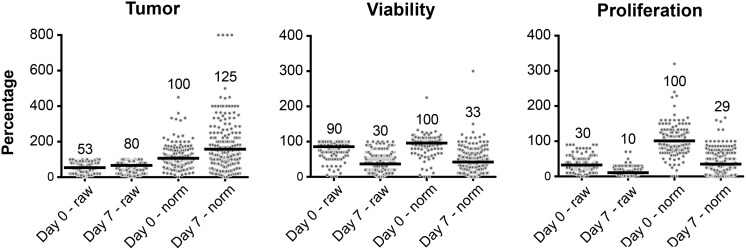
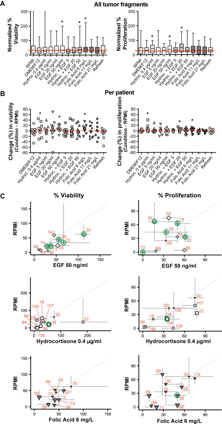
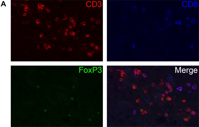
Treatment of advanced head and neck cancer is associated with low survival, high toxicity and a widely divergent individual response. The sponge-gel-supported histoculture model was previously developed to serve as a preclinical model for predicting individual treatment responses. We aimed to optimize the sponge-gel-supported histoculture model and provide more insight in cell specific behaviour by evaluating the tumor and its microenvironment using immunohistochemistry. We collected fresh tumor biopsies from 72 untreated patients and cultured them for 7 days. Biopsies from 57 patients (79%) were successfully cultured and 1451 tumor fragments (95.4%) were evaluated. Fragments were scored for percentage of tumor, tumor viability and proliferation, EGF-receptor expression and presence of T-cells and macrophages. Median tumor percentage increased from 53% at day 0 to 80% at day 7. Viability and proliferation decreased after 7 days, from 90% to 30% and from 30% to 10%, respectively. Addition of EGF, folic acid and hydrocortisone can lead to improved viability and proliferation, however this was not systematically observed. No patient subgroup could be identified with higher culture success rates. Immune cells were still present at day 7, illustrating that the tumor microenvironment is sustained. EGF supplementation did not increase viability and proliferation in patients overexpressing EGF-Receptor.
晚期头颈癌的治疗与低生存率、高毒性以及个体反应差异大有关。海绵凝胶支持的组织培养模型先前已被开发出来,用作预测个体治疗反应的临床前模型。我们旨在优化海绵凝胶支持的组织培养模型,并通过使用免疫组织化学评估肿瘤及其微环境,更深入地了解细胞的特定行为。我们从72名未经治疗的患者中收集了新鲜肿瘤活检样本,并将其培养7天。来自57名患者(79%)的活检样本成功培养,共评估了1451个肿瘤碎片(95.4%)。对碎片的肿瘤百分比、肿瘤活力和增殖、表皮生长因子受体表达以及T细胞和巨噬细胞的存在情况进行评分。肿瘤百分比中位数从第0天的53%增加到第7天的80%。7天后活力和增殖分别从90%降至30%和从30%降至10%。添加表皮生长因子、叶酸和氢化可的松可提高活力和增殖,但并非系统观察到这种情况。未发现培养成功率较高的患者亚组。免疫细胞在第7天仍然存在,这说明肿瘤微环境得以维持。在表皮生长因子受体过表达的患者中,补充表皮生长因子并未提高活力和增殖。