Lohöfer Fabian, Hoffmann Laura, Buchholz Rebecca, Huber Katharina, Glinzer Almut, Kosanke Katja, Feuchtinger Annette, Aichler Michaela, Feuerecker Benedikt, Kaissis Georgios, Rummeny Ernst J, Höltke Carsten, Faber Cornelius, Schilling Franz, Botnar René M, Walch Axel K, Karst Uwe, Wildgruber Moritz
Department of Diagnostic and Interventional Radiology, Klinikum Rechts der Isar, Technische Universität München, Munich, Germany.
Department of Analytical Chemistry, Westfälische Wilhelms Universität, Münster, Germany.
Heliyon. 2018 Apr 16;4(4):e00606. doi: 10.1016/j.heliyon.2018.e00606. eCollection 2018 Apr.
Molecular MRI is becoming increasingly important for preclinical research. Validation of targeted gadolinium probes in tissue however has been cumbersome up to now. Novel methodology to assess gadolinium distribution in tissue after in vivo application is therefore needed.
To establish combined Magnetic Resonance Imaging (MRI) and Mass Spectrometry Imaging (MSI) for improved detection and quantification of Gadofluorine P deposition in scar formation and myocardial remodeling.
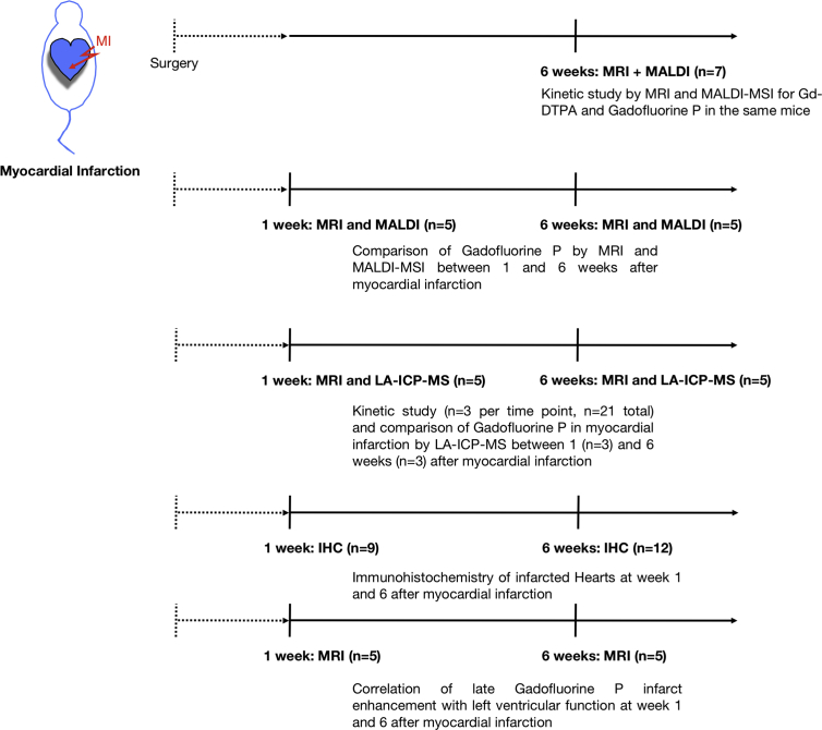
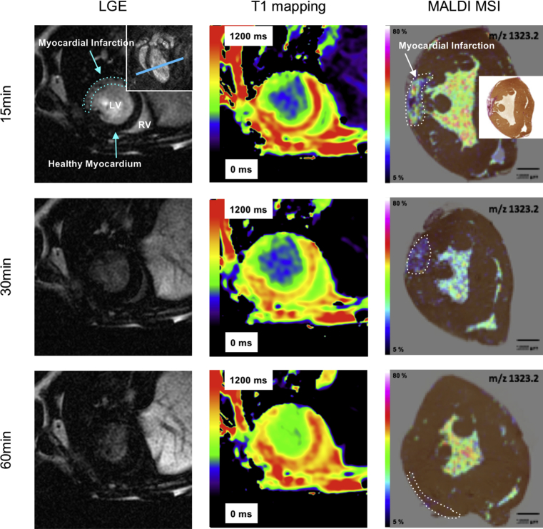
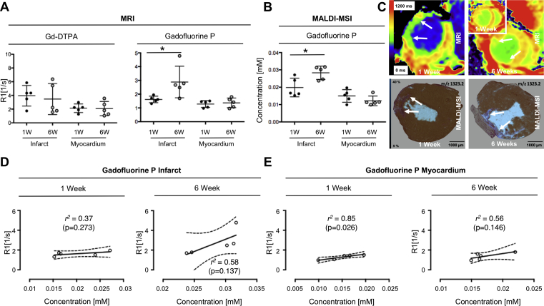
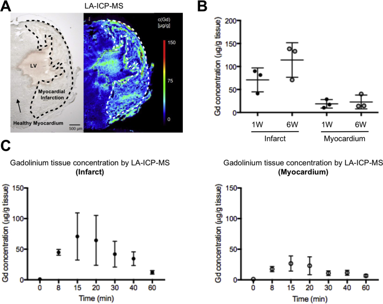
Animal studies were performed according to institutionally approved protocols. Myocardial infarction was induced by permanent ligation of the left ascending artery (LAD) in C57BL/6J mice. MRI was performed at 7T at 1 week and 6 weeks after myocardial infarction. Gadofluorine P was used for dynamic T mapping of extracellular matrix synthesis during myocardial healing and compared to Gd-DTPA. After in vivo imaging contrast agent concentration as well as distribution in tissue were validated and quantified by spatially resolved Matrix-Assisted Laser Desorption Ionization (MALDI) MSI and Laser Ablation - Inductively Coupled Plasma - Mass Spectrometry (LA-ICP-MS) imaging.
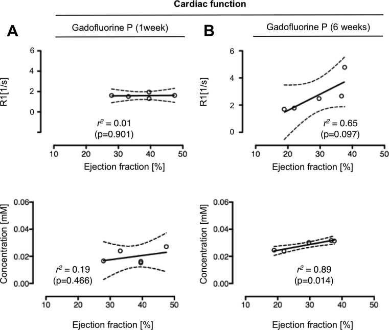
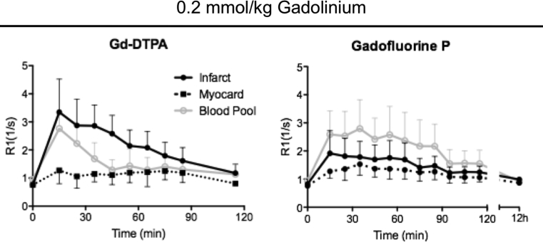
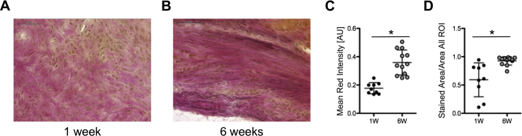
Both Gadofluorine P enhancement as well as local tissue content in the myocardial scar were highest at 15 minutes post injection. R values increased from 1 to 6 weeks after MI (1.62 s vs 2.68 s, p = 0.059) paralleled by an increase in Gadofluorine P concentration in the infarct from 0.019 mM at 1 week to 0.028 mM at 6 weeks (p = 0.048), whereas Gd-DTPA enhancement showed no differences (3.95 s vs 3.47 s, p = 0.701). MALDI-MSI results were corroborated by elemental LA-ICP-MS of Gadolinium in healthy and infarcted myocardium. Histology confirmed increased extracellular matrix synthesis at 6 weeks compared to 1 week.
Adding quantitative MSI to MR imaging enables a quantitative validation of Gadofluorine P distribution in the heart after MI for molecular imaging.
分子磁共振成像在临床前研究中变得越来越重要。然而,迄今为止,在组织中验证靶向钆探针一直很麻烦。因此,需要新的方法来评估体内应用后钆在组织中的分布。
建立联合磁共振成像(MRI)和质谱成像(MSI),以改进对钆氟磷在瘢痕形成和心肌重塑中的沉积的检测和定量。
动物研究按照机构批准的方案进行。通过永久性结扎C57BL/6J小鼠的左冠状动脉前降支(LAD)诱导心肌梗死。在心肌梗死后1周和6周时,在7T下进行MRI检查。钆氟磷用于心肌愈合过程中细胞外基质合成的动态T映射,并与钆喷酸葡胺进行比较。在体内成像后,通过空间分辨基质辅助激光解吸电离(MALDI)MSI和激光烧蚀-电感耦合等离子体质谱(LA-ICP-MS)成像对造影剂在组织中的浓度和分布进行验证和定量。
注射后15分钟时,心肌瘢痕中的钆氟磷增强以及局部组织含量均最高。心肌梗死后1至6周,R值增加(1.62秒对2.68秒,p = 0.059),梗死灶中钆氟磷浓度从1周时的0.019 mM增加至6周时的0.028 mM(p = 0.048),而钆喷酸葡胺增强无差异(3.95秒对3.47秒,p = 0.701)。健康心肌和梗死心肌中钆的元素LA-ICP-MS结果证实了MALDI-MSI结果。组织学证实,与1周相比,6周时细胞外基质合成增加。
将定量MSI添加到MR成像中,能够对心肌梗死后心脏中钆氟磷的分布进行定量验证,用于分子成像。