Department of Radiology, Stanford University School of Medicine, 300 Pasteur Drive, Stanford, CA, 94035, USA.
Division of Cardiovascular Imaging, Department of Radiology and Radiological Science, Medical University of South Carolina, Charleston, SC, USA.
Eur Radiol Exp. 2021 Mar 25;5(1):12. doi: 10.1186/s41747-021-00207-3.
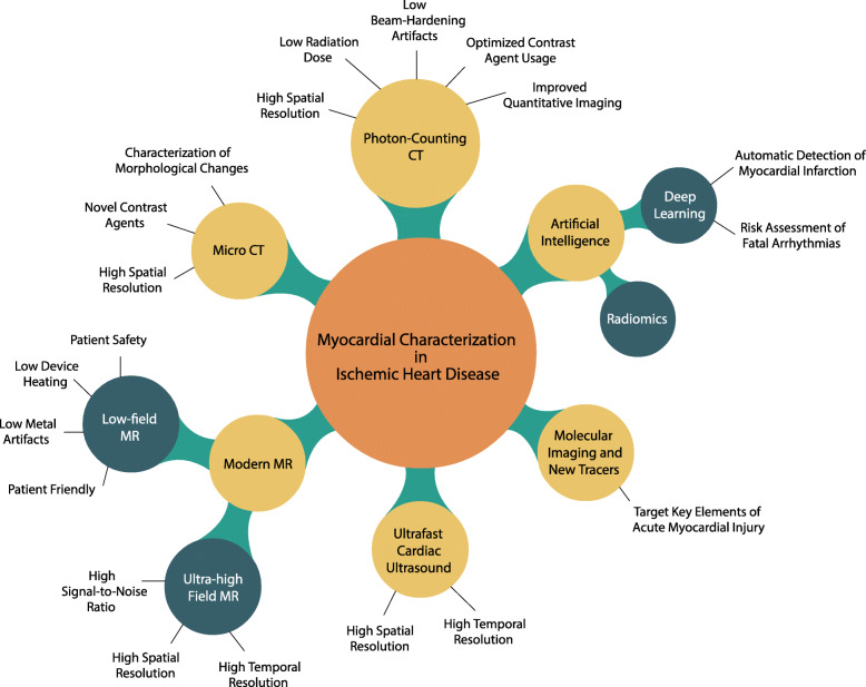
After an ischemic event, disruptive changes in the healthy myocardium may gradually develop and may ultimately turn into fibrotic scar. While these structural changes have been described by conventional imaging modalities mostly on a macroscopic scale-i.e., late gadolinium enhancement at magnetic resonance imaging (MRI)-in recent years, novel imaging methods have shown the potential to unveil an even more detailed picture of the postischemic myocardial phenomena. These new methods may bring advances in the understanding of ischemic heart disease with potential major changes in the current clinical practice. In this review article, we provide an overview of the emerging methods for the non-invasive characterization of ischemic heart disease, including coronary ultrafast Doppler angiography, photon-counting computed tomography (CT), micro-CT (for preclinical studies), low-field and ultrahigh-field MRI, and C-methionine positron emission tomography. In addition, we discuss new opportunities brought by artificial intelligence, while addressing promising future scenarios and the challenges for the application of artificial intelligence in the field of cardiac imaging.
在缺血事件发生后,健康心肌可能会逐渐发生破坏性变化,并最终转变为纤维瘢痕。虽然这些结构变化已被传统的成像方式在宏观尺度上进行了描述,即磁共振成像(MRI)中的钆延迟增强,但近年来,新的成像方法显示出揭示更详细的缺血性心肌现象的潜力。这些新方法可能会推动对缺血性心脏病的理解取得进展,并有可能在当前的临床实践中带来重大变化。在这篇综述文章中,我们提供了一种非侵入性的缺血性心脏病特征描述的新兴方法概述,包括冠状动脉超快多普勒血管造影、光子计数计算机断层扫描(CT)、微 CT(用于临床前研究)、低场和超高场 MRI 以及 C-蛋氨酸正电子发射断层扫描。此外,我们还讨论了人工智能带来的新机遇,同时探讨了人工智能在心脏成像领域应用的有前景的未来场景和挑战。