Poojan Shiv, Kim Han-Seong, Yoon Ji-Woon, Sim Hye Won, Hong Kyeong-Man
Omics Core Lab, Research Institute, National Cancer Center.
Department of Pathology, Inje University Ilsan Paik Hospital.
J Vis Exp. 2018 May 20(135):57369. doi: 10.3791/57369.
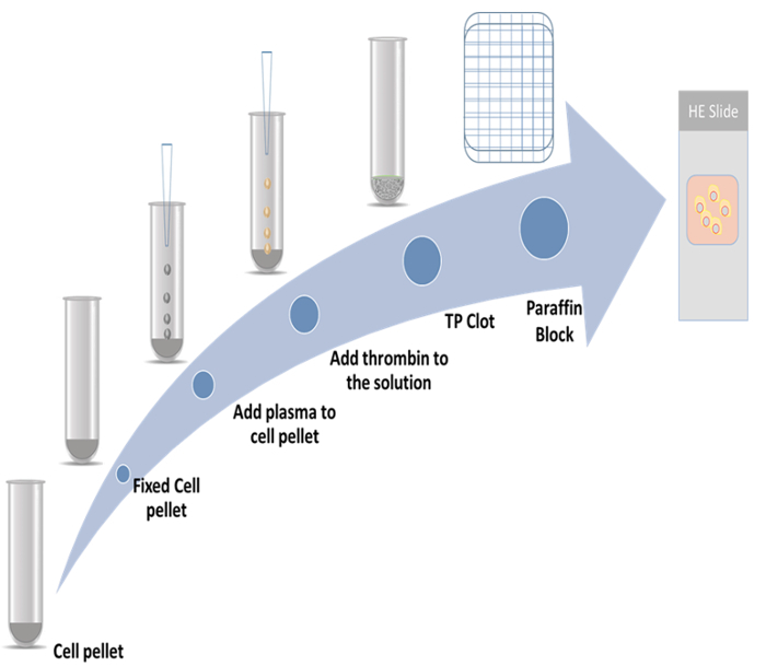
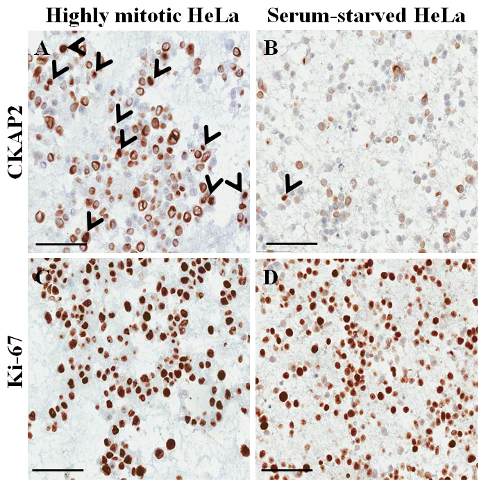
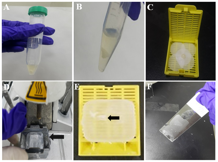
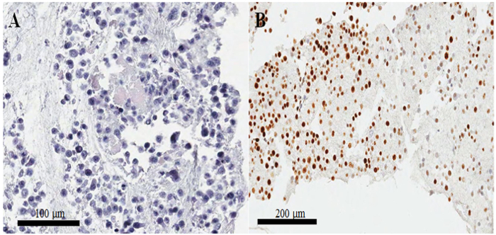
Immunofluorescent staining is currently the method of choice for determination of protein expression levels in cell-culture systems when morphological information is also necessary. The protocol of immunocytochemical staining on paraffin-embedded cell blocks, presented herein, is an excellent alternative to immunofluorescent staining on non-paraffin-embedded fixed cells. In this protocol, a paraffin cell block from HeLa cells was prepared using the thromboplastin-plasma method, and immunocytochemistry was performed for the evaluation of two proliferation markers, CKAP2 and Ki-67. The nuclei and cytoplasmic morphology of the HeLa cells were well preserved in the cell-block slides. At the same time, the CKAP2 and Ki-67 staining patterns in the immunocytochemistry were quite similar to those in immunohistochemical staining in paraffin cancer tissues. With modified cell-culture conditions, including pre-incubation of HeLa cells under serum-free conditions, the effect could be evaluated while preserving architectural information. In conclusion, immunocytochemistry on paraffin-embedded cell blocks is an excellent alternative to immunofluorescent staining.
当还需要形态学信息时,免疫荧光染色是目前在细胞培养系统中测定蛋白质表达水平的首选方法。本文介绍的石蜡包埋细胞块免疫细胞化学染色方案,是对非石蜡包埋固定细胞进行免疫荧光染色的绝佳替代方法。在该方案中,采用凝血酶原 - 血浆法制备了来自HeLa细胞的石蜡细胞块,并进行免疫细胞化学以评估两种增殖标志物CKAP2和Ki-67。HeLa细胞的细胞核和细胞质形态在细胞块载玻片上保存良好。同时,免疫细胞化学中CKAP2和Ki-67的染色模式与石蜡癌组织免疫组织化学染色中的模式非常相似。通过改良细胞培养条件,包括在无血清条件下对HeLa细胞进行预孵育,可以在保留结构信息的同时评估效果。总之,石蜡包埋细胞块的免疫细胞化学是免疫荧光染色的绝佳替代方法。