Dementia Research Centre, Department of Neurodegenerative Disease, Institute of Neurology, University College London, London, United Kingdom.
Translational Imaging Group, Centre for Medical Image Computing, University College London, London, United Kingdom.
Neuroimage Clin. 2018 Feb 23;18:675-681. doi: 10.1016/j.nicl.2018.02.019. eCollection 2018.
Frontotemporal dementia (FTD) is a heterogeneous neurodegenerative disorder associated with frontal and temporal atrophy. Subcortical involvement has been described as well, with early thalamic atrophy most commonly associated with the expansion. However thalamic involvement has not been comprehensively investigated across the FTD spectrum.
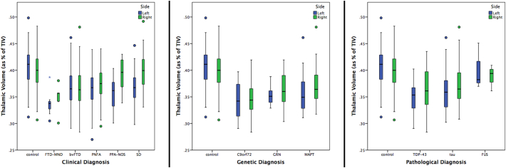
We investigated thalamic volumes in a sample of 341 FTD patients (age: mean(standard deviation) 64.2(8.5) years; disease duration: 4.6(2.7) years) compared with 99 age-matched controls (age: 61.9(11.4) years). We performed a parcellation of T1 MRIs using an atlas propagation and label fusion approach to extract left and right thalamus volumes, which were corrected for total intracranial volumes. We assessed subgroups stratified by clinical diagnosis (141 behavioural variant FTD (bvFTD), 76 semantic dementia (SD), 103 progressive nonfluent aphasia (PNFA), 7 with associated motor neurone disease (FTD-MND) and 14 primary progressive aphasia not otherwise specified (PPA-NOS), genetic diagnosis (24 with , 24 with , and 15 with mutations), and pathological diagnosis (40 tauopathy, 61 TDP-43opathy, 3 FUSopathy). We assessed the diagnostic accuracy based on thalamic volume.
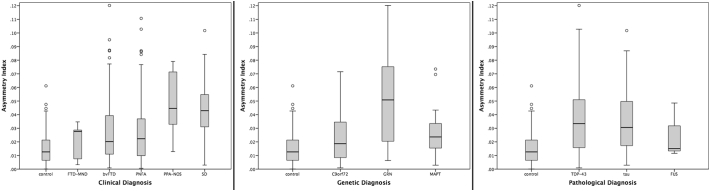
Overall, FTD patients had smaller thalami than controls (8% difference in volume, p < 0.0005, ANCOVA). Stratifying by genetics, group had the smallest thalami (14% difference from controls, p < 0.0005). However, the thalami were also smaller than controls in the other genetic groups: and groups showed a difference of 11% and 9% respectively (p < 0.0005). ROC analysis showed a relatively poor ability to separate from (AUC = 0.651, p = 0.073) and from cases (AUC = 0.644, p = 0.133) using thalamic volume. All clinical subtypes had significantly smaller thalami than controls (p < 0.0005), with the FTD-MND group having the smallest (15%), followed by bvFTD (9%), PNFA (8%), PPA-NOS (7%), and lastly SD (5%). In the pathological groups, the TDP-43opathies had an 11% difference from controls, and tauopathies 9%, while the FUSopathies showed only 2% of difference from controls (p < 0.0005). , PPA-NOS and SD were the subgroups showing the highest asymmetry in volumes.
The thalamus was most affected in genetically, TDP-43opathies pathologically and FTD-MND clinically. However, thalamic atrophy is a common feature across all FTD groups.
额颞叶痴呆(FTD)是一种与额颞叶萎缩相关的异质性神经退行性疾病。也有描述涉及皮质下区域,最常见的与扩展相关的是丘脑早期萎缩。然而,FTD 谱中尚未全面研究丘脑的参与情况。
我们在 341 名 FTD 患者(年龄:平均值(标准差)64.2(8.5)岁;疾病持续时间:4.6(2.7)年)与 99 名年龄匹配的对照组(年龄:61.9(11.4)岁)中研究了丘脑体积。我们使用图谱传播和标签融合方法对 T1 MRI 进行了分割,以提取左右丘脑体积,并对其进行了总颅内体积校正。我们根据临床诊断进行了亚组评估(141 例行为变异型 FTD(bvFTD)、76 例语义性痴呆(SD)、103 例进行性非流利性失语症(PNFA)、7 例伴有运动神经元病的 FTD(FTD-MND)和 14 例非特定的原发性进行性失语症(PPA-NOS)、遗传诊断(24 例携带、24 例携带、15 例携带突变)和病理诊断(40 例 tau 病变、61 例 TDP-43 病变、3 例 FUS 病变)。我们基于丘脑体积评估了诊断准确性。
总体而言,FTD 患者的丘脑体积比对照组小(体积差异 8%,p<0.0005,ANCOVA)。按遗传学分层,组的丘脑最小(与对照组相差 14%,p<0.0005)。然而,其他遗传组的丘脑也比对照组小:和组分别显示出 11%和 9%的差异(p<0.0005)。ROC 分析显示,使用丘脑体积区分与的能力相对较差(AUC=0.651,p=0.073),与病例的区分能力也较差(AUC=0.644,p=0.133)。所有临床亚型的丘脑体积均明显小于对照组(p<0.0005),FTD-MND 组最小(15%),其次是 bvFTD(9%)、PNFA(8%)、PPA-NOS(7%)和 SD(5%)。在病理组中,TDP-43 病变与对照组相比有 11%的差异,tau 病变有 9%的差异,而 FUS 病变与对照组相比只有 2%的差异(p<0.0005)。、PPA-NOS 和 SD 是体积差异最大的亚组。
在遗传学上,TDP-43 病变在病理学上和 FTD-MND 在临床上丘脑受影响最大。然而,丘脑萎缩是所有 FTD 组的共同特征。